. 2015 Mar 27:15:18.
doi: 10.1186/s12861-015-0066-9.

Development and origins of zebrafish ocular vasculature

Affiliations

Development and origins of zebrafish ocular vasculature

Rivka Kaufman et al. BMC Dev Biol. .

Abstract

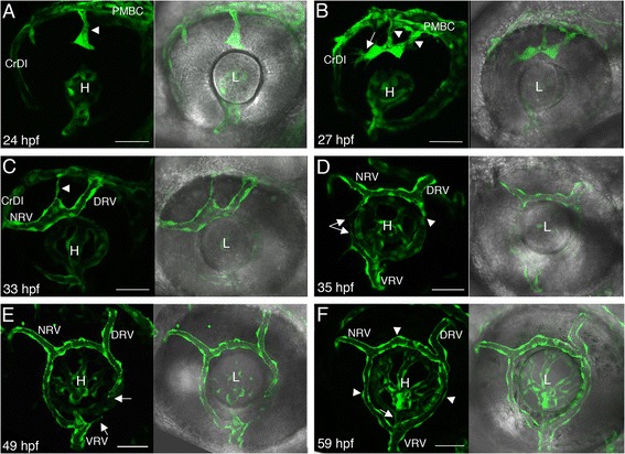
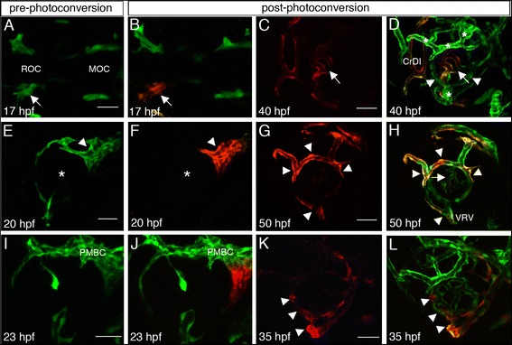
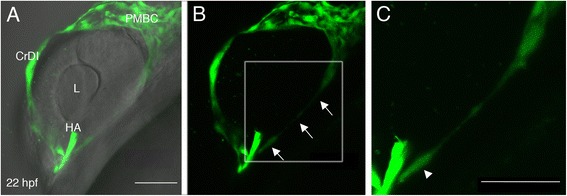
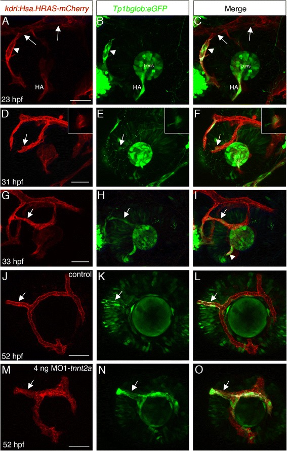
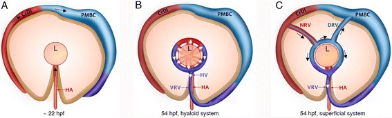
Background: The developing eye receives blood supply from two vascular systems, the intraocular hyaloid system and the superficial choroidal vessels. In zebrafish, a highly stereotypic and simple set of vessels develops on the surface of the eye prior to development of choroidal vessels. The origins and formation of this so-called superficial system have not been described.

Results: We have analyzed the development of superficial vessels by time-lapse imaging and identified their origins by photoconversion experiments in kdrl:Kaede transgenic embryos. We show that the entire superficial system is derived from a venous origin, and surprisingly, we find that the hyaloid system has, in addition to its previously described arterial origin, a venous origin for specific vessels. Despite arising solely from a vein, one of the vessels in the superficial system, the nasal radial vessel (NRV), appears to acquire an arterial identity while growing over the nasal aspect of the eye and this happens in a blood flow-independent manner.

Conclusions: Our results provide a thorough analysis of the early development and origins of zebrafish ocular vessels and establish the superficial vasculature as a model for studying vascular patterning in the context of the developing eye.


Figures

Figure 1
Figure 1
Development of the superficial system. (A-F) Projections of confocal z-stacks (left in each panel) showing ocular vessels in Tg(kdrl:EGFP) live embryos at different developmental time points, which are depicted in each panel. On the right side of each panel are the same conofocal images combined with bright field images showing position of vessels relative to eye tissues. (A) Arrowhead points at an initial sprout that will form the DRV. (B) Arrowheads point at two vessels, one or both will form the DRV. These vessels have connected and the tip cell (arrow) grows towards the CrDI. (C) The DRV and NRV have formed. One of the two initial vessels that sprouted from the PMBC is being pruned (arrowhead). (D) Sprouts arising from the VRV and NRV/SAV junction send long filopodial extensions towards each other (arrows). The posterior part of the SAV begins to grow ventrally (arrowhead). (E) The posterior SAV continues to grow ventrally, but there is only minor angiogenic activity from the VRV. Arrows point at tip cell of posterior SAV and small filopodial extension from the VRV. (F) The completed superficial system. Arrow points at the hyaloid vein and arrowheads at the SAV. All images are lateral views, anterior to the left, dorsal up. CrDI, cranial division of internal carotid artery; DRV, dorsal retinal vessel; H, hyaloid system; L, lens; NRV, nasal radial vessel; PMBC, Primordial midbrain channel; SAV, superficial annular vessel; VRV, ventral radial vessel. Scale bars are 50 μm.
Figure 2
Figure 2
Origins of superficial and hyaloid vessels. (A,E,I) Pre-photoconversion and (B-D, F-H, J-L) post-photoconversion confocal z-stack projections of kdrl:Kaede embryos. Cells that were not photoconverteed are green whereas photoconverted cells are red. Age of embryos when imaged is depicted in each panel. (A-D) Photoconversion of left ROC. (A,B) Arrows point at the left ROC. (C,D) Single channel image showing only photoconverted endothelial cells (C), and a merge of channels showing photoconverted and non-photoconverted cells (D). Arrows and arrowheads point at central hyaloid vessels with photoconverted cells and more peripheral hyaloid vessels without photoconverted cells, respectively. (E-H) Photoconversion of PMBC. (E,F) Asterisks mark the center of the eye. (G,H) Single channel image showing only photoconverted endothelial cells (G), and a merge of channels showing photoconverted and non-photoconverted cells (H). Arrowheads point at superficial vessels and arrow in H points at hyaloid vessels. (I-L) Photoconversion of ventral PMBC. (I,J) Only the ventral region of the PMBC was photoconverted (red in J). (K,L) Single channel image showing only photoconverted endothelial cells (K), and a merge of channels showing photoconverted and non-photoconverted cells (L). Arrowheads point at the VRV and peripheral hyaloid vessels. (A,B) are dorsal views, all other panels are lateral views, anterior to the left. CrDI, cranial division of internal carotid artery; DRV, dorsal retinal vessel; MOC, midbrain organizing center; NRV, nasal radial vessel; PMBC, Primordial midbrain channel; ROC, rostral organizing center; VRV, ventral radial vessel. Scale bars are 50 μm.
Figure 3
Figure 3
Sprouting from the ventral PMBC. (A-C) Projections of confocal z-stacks from a live kdrl:EGFP transgenic embryo at 22 hpf. In A, the same confocal image shown in B is combined with bright field image to demonstrate position of blood vessels relative to eye tissues. Arrows in B point at a long extension from the ventral PMBC towards the optic fissure, where the HA enters the eye. (C) A higher magnification of the region marked by white rectangle in B suggests the extension is actually a trail of cells. Arrowhead in C points at what appears to be an endothelial cell body. CrDI, cranial division of internal carotid artery; HA, hyaloid artery; L, lens; PMBC, Primordial midbrain channel. Anterior is to the left and dorsal up. Scale bars are 50 μm.
Figure 4
Figure 4
Notch pathway activation in the NRV. (A-O) Single channel or merged confocal z-stack projections of double transgenic embryos carrying kdrl:Hsa.HRAS-mCherry (red) and Tp1bglob:eGFP (green) transgenes. (A-C) At 23 hpf, the CrDI (arrowhead) expresses EGFP whereas the PMBC (arrows in A and C) does not. (D-I) At 31–33 hpf the NRV is close to connecting to the CrDI (D-F) or has already connected (G-I). EGFP expression can be seen in one of the leading cells of the NRV (arrows in D-F). Insets are higher magnification of the region the arrows point at and are 4 μm single confocal sections. EGFP expression becomes clearer throughout the NRV once it is connected to the CrDI (arrows in G-I). (J-O) Control (J-L) and MO1-tnnt2a injected (M-O) embryos at approximately 52 hpf. EGFP expression in the NRV (arrows) is evident in injected embryos. HA, hyaloid artery. All images are lateral views, anterior to the left, dorsal up. CrDI, cranial division of internal carotid artery; DRV, dorsal retinal vessel; HA, hyaloid artery; NRV, nasal radial vessel; PMBC, Primordial midbrain channel; VRV, ventral radial vessel. Scale bars are 50 μm.
Figure 5
Figure 5
Summary of ocular vessel origins. (A-C) Schematic illustration of ocular vessel origin. The colors of vessels represent their origins: from the dorsal PMBC - light blue, ventral PMBC - purple and from the HA – red (excluding the CrDI). Blue and purple represent venous identity, red represents arterial identity. (A) Just before superficial vessels grow, the PMBC and CrDI have connected and the HA entered the eye through the optic fissure. (B) The hyaloid system. Vessels are located inside the eye, at a deeper level than vessels shown in C. (C) The completed superficial system. The NRV is red, indicating its arterial identity with a blue core representing its origin from the dorsal PMBC. Arrows in B and C show direction of blood flow. CrDI, cranial division of internal carotid artery; DRV, dorsal retinal vessel; HA, hyaloid artery; HV, hyaloid vein; L, lens; NRV, nasal radial vessel; PMBC, Primordial midbrain channel; SAV, superficial annular vessel; VRV, ventral radial vessel.

References

    1. Heinke J, Patterson C, Moser M. Life is a pattern: vascular assembly within the embryo. Front Biosci (Elite Ed) 2012;4:2269–88. doi: 10.2741/E541. - DOI - PMC - PubMed
    1. Ellertsdottir E, Lenard A, Blum Y, Krudewig A, Herwig L, Affolter M, et al. Vascular morphogenesis in the zebrafish embryo. Dev Biol. 2010;341:56–65. doi: 10.1016/j.ydbio.2009.10.035. - DOI - PubMed
    1. Saint-Geniez M, D’Amore PA. Development and pathology of the hyaloid, choroidal and retinal vasculature. Int J Dev Biol. 2004;48:1045–58. doi: 10.1387/ijdb.041895ms. - DOI - PubMed
    1. Alvarez Y, Cederlund ML, Cottell DC, Bill BR, Ekker SC, Torres-Vazquez J, et al. Genetic determinants of hyaloid and retinal vasculature in zebrafish. BMC Dev Biol. 2007;7:114. doi: 10.1186/1471-213X-7-114. - DOI - PMC - PubMed
    1. Hartsock A, Lee C, Arnold V, Gross JM. In vivo analysis of hyaloid vasculature morphogenesis in zebrafish: a role for the lens in maturation and maintenance of the hyaloid. Dev Biol. 2014;394:327–39. doi: 10.1016/j.ydbio.2014.07.024. - DOI - PMC - PubMed

Publication types

LinkOut - more resources