. 2015 Jun 15;308(12):H1530-9.
doi: 10.1152/ajpheart.00008.2015. Epub 2015 Apr 17.

Cardiac mTOR rescues the detrimental effects of diet-induced obesity in the heart after ischemia-reperfusion

Affiliations

Cardiac mTOR rescues the detrimental effects of diet-induced obesity in the heart after ischemia-reperfusion

Toshinori Aoyagi et al. Am J Physiol Heart Circ Physiol. .

Abstract

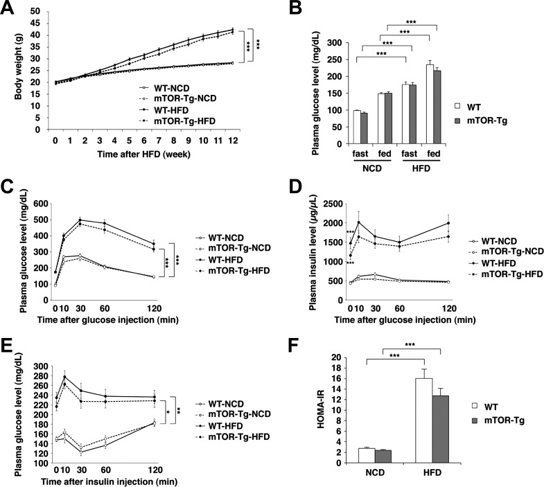
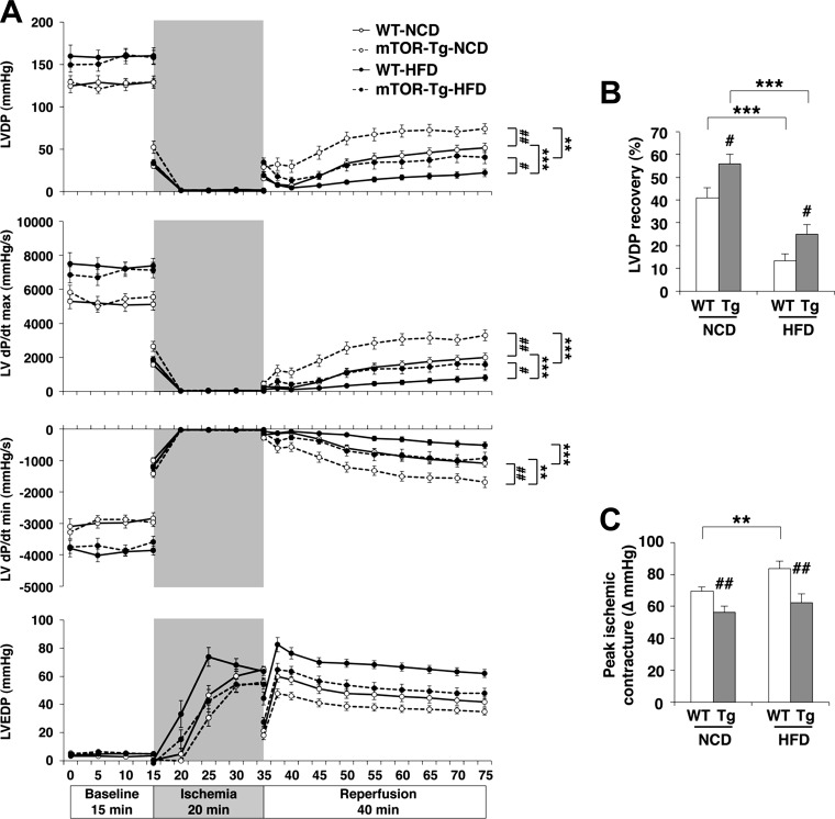
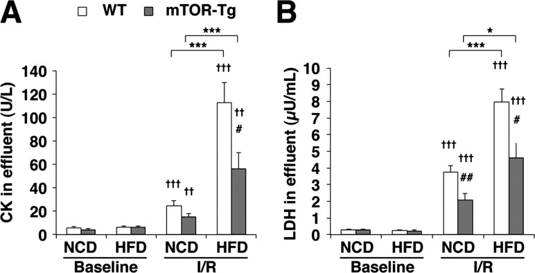
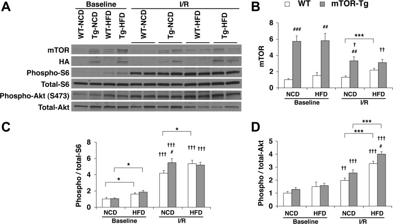
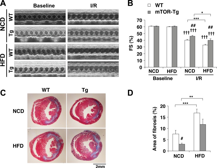
Diet-induced obesity deteriorates the recovery of cardiac function after ischemia-reperfusion (I/R) injury. While mechanistic target of rapamycin (mTOR) is a key mediator of energy metabolism, the effects of cardiac mTOR in ischemic injury under metabolic syndrome remains undefined. Using cardiac-specific transgenic mice overexpressing mTOR (mTOR-Tg mice), we studied the effect of mTOR on cardiac function in both ex vivo and in vivo models of I/R injury in high-fat diet (HFD)-induced obese mice. mTOR-Tg and wild-type (WT) mice were fed a HFD (60% fat by calories) for 12 wk. Glucose intolerance and insulin resistance induced by the HFD were comparable between WT HFD-fed and mTOR-Tg HFD-fed mice. Functional recovery after I/R in the ex vivo Langendorff perfusion model was significantly lower in HFD-fed mice than normal chow diet-fed mice. mTOR-Tg mice demonstrated better cardiac function recovery and had less of the necrotic markers creatine kinase and lactate dehydrogenase in both feeding conditions. Additionally, mTOR overexpression suppressed expression of proinflammatory cytokines, including IL-6 and TNF-α, in both feeding conditions after I/R injury. In vivo I/R models showed that at 1 wk after I/R, HFD-fed mice exhibited worse cardiac function and larger myocardial scarring along myofibers compared with normal chow diet-fed mice. In both feeding conditions, mTOR overexpression preserved cardiac function and prevented myocardial scarring. These findings suggest that cardiac mTOR overexpression is sufficient to prevent the detrimental effects of diet-induced obesity on the heart after I/R, by reducing cardiac dysfunction and myocardial scarring.

Keywords: diet-induced obesity; heart failure; mammalian target of rapamycin; metabolic syndrome; myocardial infarction; transgenic mice.


Figures

Fig. 1.
Fig. 1.
High-fat diet (HFD)-induced obesity, glucose intolerance, and insulin resistance are comparable between wild-type (WT) and mechanistic target of rapamycin (mTOR)-overexpressing transgenic (mTOR-Tg) mice. A: body weight changes during HFD feeding (n = 36–40 mice/group). B: 16-h fasting or fed plasma glucose levels [n = 24 WT mice fed a normal chow diet (WT-NCD mice), 24 mTOR-Tg fed a NCD (mTOR-Tg-NCD mice), 22 WT mice fed a HFD (WT-HFD mice), and 22 mTOR-Tg mice fed a HFD (mTOR-Tg-HFD mice)]. C and D: plasma glucose and insulin levels during glucose tolerance tests. E: plasma glucose levels during insulin tolerance tests. F: homeostatic model of assessment of insulin resistance (HOMA-IR) calculated with the formula described in materials and methods. *P < 0.05, **P < 0.01, and ***P < 0.001, HFD vs. NCD (by two-way ANOVA or Student's t-test).
Fig. 2.
Fig. 2.
Overexpression of cardiac mTOR prevents cardiac dysfunction after transient ischemia in HFD hearts. A: left ventricular (LV) developed pressure (LVDP), LV dP/dtmax, LV dP/dtmin, and LV end-diastolic pressure (LVEDP) during I/R in WT-NCD, mTOR-Tg-NCD, WT-HFD, and mTOR-Tg-NCD hearts. B: maximum LVDP recovery (percentage of baseline) measured at 40 min of reperfusion. C: ischemic contracture during the 20-min ischemia period as determined by peak ischemic contracture (ΔLVEDP from 0 min of ischemia). n = 24 WT-NCD mice, 28 mTOR-Tg-NCD mice, 24 WT-HFD mice, and 26 mTOR-Tg-HFD mice. *P < 0.05, **P < 0.01, and ***P < 0.001, NCD vs. HFD; #P < 0.05 and ##P < 0.01, WT vs. mTOR-Tg mice (by two-way ANOVA or Student's t-test).
Fig. 3.
Fig. 3.
Overexpression of cardiac mTOR prevents cardiac injury after ex vivo transient ischemia in HFD hearts. A and B: activities of creatine kinase (CK; A) and lactate dehydrogenase (LDH; B) in the effluent collected during the reperfusion period. To determine enzyme activities immediately after ex vivo I/R injury, effluents from hearts exposed to either 20 or 40 min of global ischemia were collected at control perfusion (baseline) and after 40-min reperfusion (I/R). n = 14 WT-NCD mice, 5 mTOR-Tg-NCD mice, 13 WT-HFD mice, 15 mTOR-Tg-HFD mice. *P < 0.05 and ***P < 0.001, NCD vs. HFD; #P < 0.05 and ##P < 0.01, WT vs. mTOR-Tg mice; ††P < 0.01 and †††P < 0.001, baseline vs. I/R (by Student's t-test).
Fig. 4.
Fig. 4.
Overexpression of cardiac mTOR induces functional activation of both mTORC1 and mTORC2 in post-I/R hearts. A: representative immunoblots of mTOR signaling molecules in hearts subjected to the ex vivo Langendorff perfusion model. Baseline hearts were harvested after 15 min of equilibration perfusion ex vivo. I/R hearts were harvested after a course of baseline conditions followed by 20-min ischemia and then 40-min reperfusion. Immunoblot analysis was performed with the indicated antibodies. Blots are representative of six independent experiments. Densitometric quantitative analyses of mTOR (B), phospho-S6 (C), and phospho-Akt (D) were normalized to baseline levels of WT-NCD hearts in each experiment. n = 6 baseline hearts and 12 I/R hearts. *P < 0.05 and ***P < 0.001, NCD vs. HFD; #P < 0.05, ##P < 0.01, and ###P < 0.001, WT vs. mTOR-Tg hearts; †P < 0.05, ††P < 0.01, and †††P < 0.001, baseline vs. I/R (by Student's t-test).
Fig. 5.
Fig. 5.
Overexpression of mTOR does not affect autophagic activity in a NCD nor HFD. A: representative immunoblots of light chain 3 [LC3; LC3-I (top) and LC3-II (bottom)] and GAPDH levels in hearts subjected to the ex vivo Langendorff perfusion model. Hearts were harvested after I/R as described in Fig 4. B: densitometric analysis of LC3-II levels normalized to GAPDH. n = 6 for all groups.
Fig. 6.
Fig. 6.
Overexpression of mTOR suppresses the induction of proinflammatory cytokines and chemokines after I/R in HFD-fed hearts. mRNA expression levels of proinflammatory cytokines (IL-6, IL-1β, and TNF-α), chemokines [monocyte chemotactic protein (MCP)-1 and macrophage inflammatory protein (MIP)-1α], and growth differentiation factor (GDF)15 in hearts subjected to ex vivo I/R are shown. mRNA was measured by quantitative real-time PCR. n = 6 for each group of baseline hearts; n = 12 WT-NCD hearts, 12 mTOR-Tg-NCD hearts; 10 WT-HFD hearts, and 14 mTOR-Tg-HFD hearts for I/R. #P < 0.05 and ##P < 0.01, WT vs. mTOR-Tg hearts; ††P < 0.01 and †††P < 0.001, baseline vs. I/R (by Student's t-test).
Fig. 7.
Fig. 7.
Overexpression of mTOR increases the level of GDF15 protein at baseline. Representative immunoblots compare expression of mTOR and GDF15 in hearts isolated from WT and mTOR-Tg mice without additional treatments. Immunoblots were performed with the primary antibodies described in materials and methods. HA, hemagglutinin. Blots are representative of n = 5 WT mice and 6 mTOR-Tg mice. Densitometry graphs were normalized to GAPDH and represent normalized average densities ± SE. *P < 0.05, **P < 0.01, and ***P < 0.001 (by Student's t-test).
Fig. 8.
Fig. 8.
Overexpression of cardiac mTOR preserves cardiac function after in vivo transient ischemia in HFD hearts. A: representative M-mode images of operated WT-NCD, mTOR-Tg-NCD, WT-HFD, and mTOR-Tg-HFD mice at baseline and 1 wk after I/R surgery. B: mean scores for fractional shortening (FS; in %) at 1 wk after I/R. C: representative Masson's trichrome staining after 1 wk of surgery. D: quantitative analysis of interstitial fibrosis detected by Masson's trichrome staining. n = 12 WT-NCD hearts, 12 mTOR-Tg-NCD hearts, 15 WT-HFD hearts, and 10 mTOR-Tg-HFD hearts. *P < 0.05, **P < 0.01, and ***P < 0.001, NCD vs. HFD; #P < 0.05 and ##P < 0.01, WT vs. mTOR-Tg hearts; †††P < 0.001, baseline vs. I/R (by Student's t-test).

References

    1. Aguilar D, Solomon SD, Kober L, Rouleau JL, Skali H, McMurray JJ, Francis GS, Henis M, O'Connor CM, Diaz R, Belenkov YN, Varshavsky S, Leimberger JD, Velazquez EJ, Califf RM, Pfeffer MA. Newly diagnosed and previously known diabetes mellitus and 1-year outcomes of acute myocardial infarction: the VALsartan In Acute myocardial iNfarcTion (VALIANT) trial. Circulation 110: 1572–1578, 2004. - PubMed
    1. Alberti KG, Eckel RH, Grundy SM, Zimmet PZ, Cleeman JI, Donato KA, Fruchart JC, James WP, Loria CM, Smith SC Jr. Harmonizing the metabolic syndrome: a joint interim statement of the International Diabetes Federation Task Force on Epidemiology and Prevention; National Heart, Lung, and Blood Institute; American Heart Association; World Heart Federation; International Atherosclerosis Society; and International Association for the Study of Obesity. Circulation 120: 1640–1645, 2009. - PubMed
    1. Ameri K, Harris AL. Activating transcription factor 4. Int J Biochem Cell Biol 40: 14–21, 2008. - PubMed
    1. Aoyagi T, Birumachi J, Hiroyama M, Fujiwara Y, Sanbe A, Yamauchi J, Tanoue A. Alteration of glucose homeostasis in V1a vasopressin receptor-deficient mice. Endocrinology 148: 2075–2084, 2007. - PubMed
    1. Aoyagi T, Kusakari Y, Xiao CY, Inouye BT, Takahashi M, Scherrer-Crosbie M, Rosenzweig A, Hara K, Matsui T. Cardiac mTOR protects the heart against ischemia-reperfusion injury. Am J Physiol Heart Circ Physiol 303: H75–H85, 2012. - PMC - PubMed

Publication types

MeSH terms

LinkOut - more resources