

PC-PLC/sphingomyelin synthase activity plays a central role in the development of myogenic tone in murine resistance arteries
- PMID: 25888510
- PMCID: PMC4469871
- DOI: 10.1152/ajpheart.00594.2014
PC-PLC/sphingomyelin synthase activity plays a central role in the development of myogenic tone in murine resistance arteries
Abstract
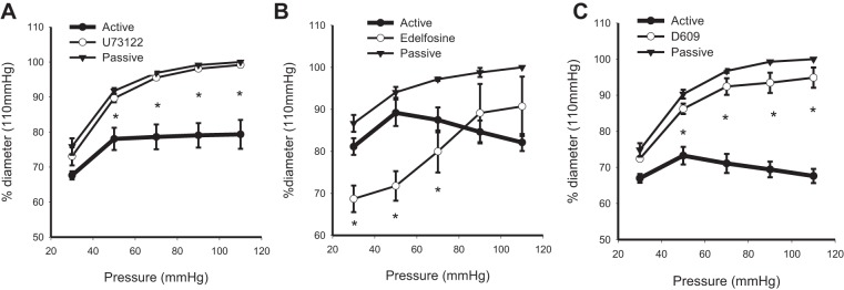
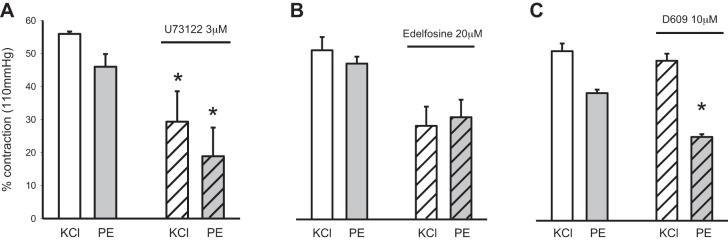
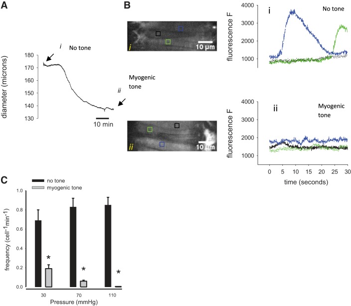
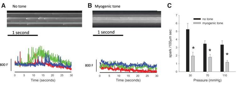
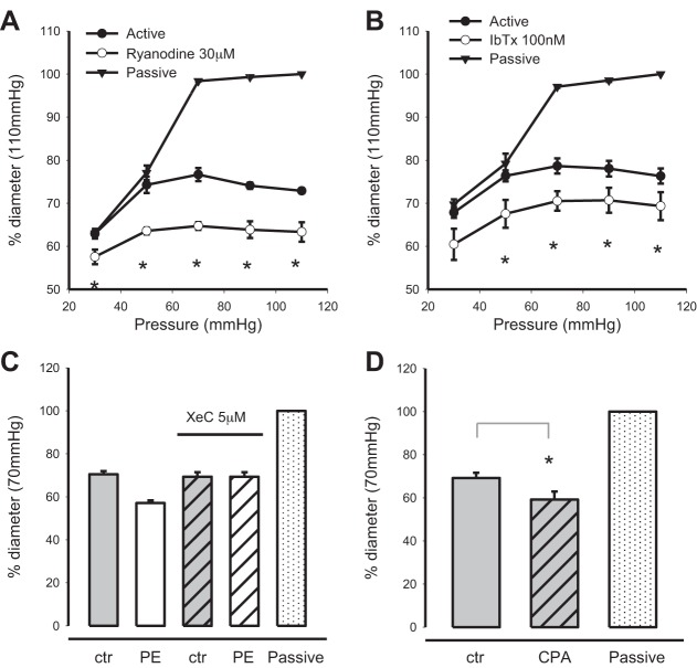
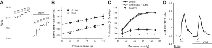
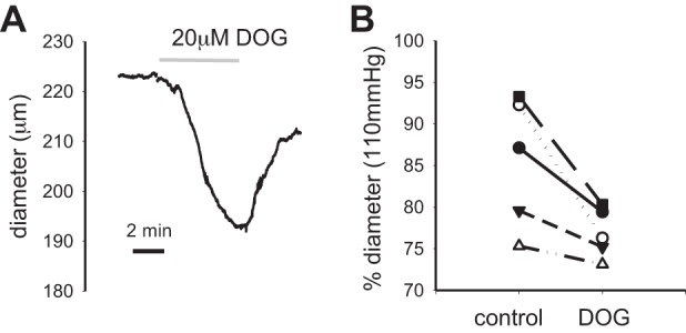
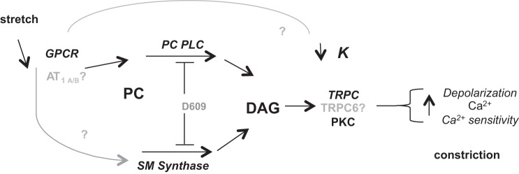
Myogenic tone is an intrinsic property of the vasculature that contributes to blood pressure control and tissue perfusion. Earlier investigations assigned a key role in myogenic tone to phospholipase C (PLC) and its products, inositol 1,4,5-trisphosphate (IP3) and diacylglycerol (DAG). Here, we used the PLC inhibitor, U-73122, and two other, specific inhibitors of PLC subtypes (PI-PLC and PC-PLC) to delineate the role of PLC in myogenic tone of pressurized murine mesenteric arteries. U-73122 inhibited depolarization-induced contractions (high external K(+) concentration), thus confirming reports of nonspecific actions of U-73122 and its limited utility for studies of myogenic tone. Edelfosine, a specific inhibitor of PI-PLC, did not affect depolarization-induced contractions but modulated myogenic tone. Because PI-PLC produces IP3, we investigated the effect of blocking IP3 receptor-mediated Ca(2+) release on myogenic tone. Incubation of arteries with xestospongin C did not affect tone, consistent with the virtual absence of Ca(2+) waves in arteries with myogenic tone. D-609, an inhibitor of PC-PLC and sphingomyelin synthase, strongly inhibited myogenic tone and had no effect on depolarization-induced contraction. D-609 appeared to act by lowering cytoplasmic Ca(2+) concentration to levels below those that activate contraction. Importantly, incubation of pressurized arteries with a membrane-permeable analog of DAG induced vasoconstriction. The results therefore mandate a reexamination of the signaling pathways activated by the Bayliss mechanism. Our results suggest that PI-PLC and IP3 are not required in maintaining myogenic tone, but DAG, produced by PC-PLC and/or SM synthase, is likely through multiple mechanisms to increase Ca(2+) entry and promote vasoconstriction.
Keywords: Bayliss; calcium; diacylglycerol; phospholipase C.
Copyright © 2015 the American Physiological Society.
Figures
Similar articles
-
Role of phospholipase C in development of myogenic tone in rat posterior cerebral arteries.Am J Physiol Heart Circ Physiol. 2002 Dec;283(6):H2234-8. doi: 10.1152/ajpheart.00624.2002. Epub 2002 Aug 29. Am J Physiol Heart Circ Physiol. 2002. PMID: 12388230
-
Divergent signaling mechanisms for venous versus arterial contraction as revealed by endothelin-1.J Vasc Surg. 2015 Sep;62(3):721-33. doi: 10.1016/j.jvs.2014.03.010. Epub 2014 Apr 14. J Vasc Surg. 2015. PMID: 24726828 Free PMC article.
-
Endothelin-1 stimulates hydrolysis of phosphatidylcholine by phospholipases C and D in intact rat mesenteric arteries.J Vasc Res. 1999 Jan-Feb;36(1):35-46. doi: 10.1159/000025624. J Vasc Res. 1999. PMID: 10050072
-
Signal transduction in esophageal and LES circular muscle contraction.Yale J Biol Med. 1999 Mar-Jun;72(2-3):153-68. Yale J Biol Med. 1999. PMID: 10780577 Free PMC article. Review.
-
Plant phospholipase C family: Regulation and functional role in lipid signaling.Cell Calcium. 2015 Aug;58(2):139-46. doi: 10.1016/j.ceca.2015.04.003. Epub 2015 Apr 17. Cell Calcium. 2015. PMID: 25933832 Review.
Cited by
-
Role of age, Rho-kinase 2 expression, and G protein-mediated signaling in the myogenic response in mouse small mesenteric arteries.Physiol Rep. 2018 Sep;6(17):e13863. doi: 10.14814/phy2.13863. Physiol Rep. 2018. PMID: 30198176 Free PMC article.
-
Calcium Channels in Vascular Smooth Muscle.Adv Pharmacol. 2017;78:49-87. doi: 10.1016/bs.apha.2016.08.002. Epub 2016 Oct 14. Adv Pharmacol. 2017. PMID: 28212803 Free PMC article. Review.
-
Smooth Muscle Ion Channels and Regulation of Vascular Tone in Resistance Arteries and Arterioles.Compr Physiol. 2017 Mar 16;7(2):485-581. doi: 10.1002/cphy.c160011. Compr Physiol. 2017. PMID: 28333380 Free PMC article. Review.
-
A Comprehensive Review on the Interplay between Neisseria spp. and Host Sphingolipid Metabolites.Cells. 2021 Nov 17;10(11):3201. doi: 10.3390/cells10113201. Cells. 2021. PMID: 34831424 Free PMC article. Review.
-
Voltage-gated Ca2+ channel activity modulates smooth muscle cell calcium waves in hamster cremaster arterioles.Am J Physiol Heart Circ Physiol. 2018 Oct 1;315(4):H871-H878. doi: 10.1152/ajpheart.00292.2018. Epub 2018 Jun 29. Am J Physiol Heart Circ Physiol. 2018. PMID: 29957015 Free PMC article.
References
-
- Amtmann E. The antiviral, antitumoural xanthate D609 is a competitive inhibitor of phosphatidylcholine-specific phospholipase C. Drugs Exp Clin Res 22: 287–294, 1996. - PubMed
-
- Bakker EN, Kerkhof CJ, Sipkema P. Signal transduction in spontaneous myogenic tone in isolated arterioles from rat skeletal muscle. Cardiovasc Res 41: 229–236, 1999. - PubMed
-
- Ben-Chaim Y, Chanda B, Dascal N, Bezanilla F, Parnas I, Parnas H. Movement of ‘gating charge’ is coupled to ligand binding in a G-protein-coupled receptor. Nature 444: 106–109, 2006. - PubMed
Publication types
MeSH terms
Substances
Grants and funding
LinkOut - more resources
Full Text Sources
Other Literature Sources
Research Materials
Miscellaneous