. 2015 May 19;84(20):2021-8.
doi: 10.1212/WNL.0000000000001591. Epub 2015 Apr 17.

The surgically remediable syndrome of epilepsy associated with bottom-of-sulcus dysplasia

Affiliations

The surgically remediable syndrome of epilepsy associated with bottom-of-sulcus dysplasia

A Simon Harvey et al. Neurology. .

Abstract

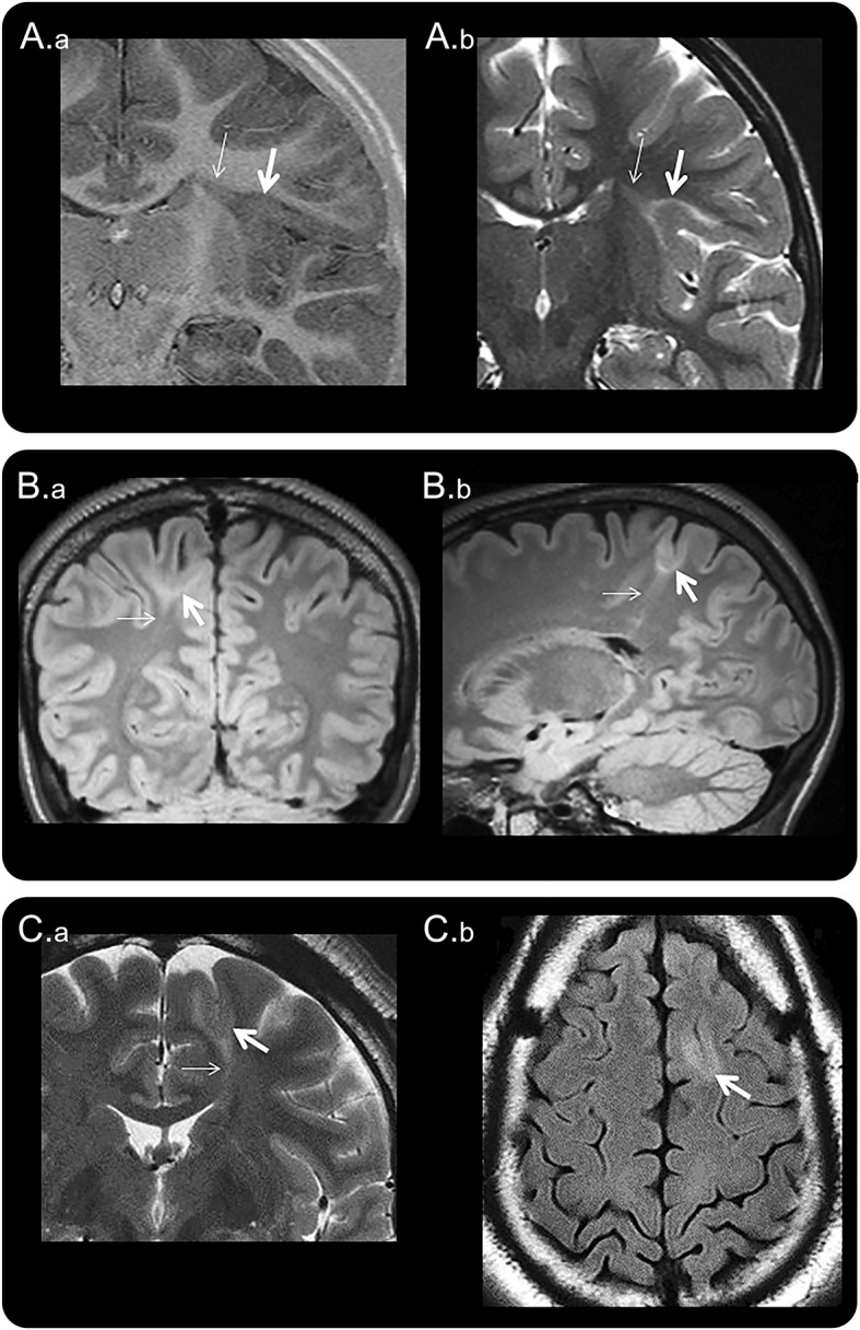
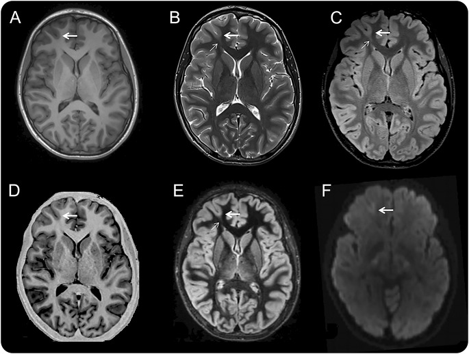
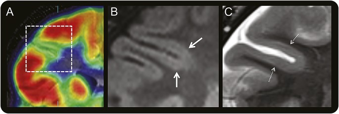
Objective: To determine clinical and EEG features that might help identify patients with epilepsy harboring small, intrinsically epileptogenic, surgically treatable, bottom-of-sulcus dysplasias (BOSDs).

Methods: Retrospective review of clinical records, EEG, MRI, and histopathology in 32 patients with drug-resistant epilepsy and MRI-positive (72% 3.0 tesla), pathologically proven (type 2B cortical dysplasia) BOSDs operated at our centers during 2005-2013.

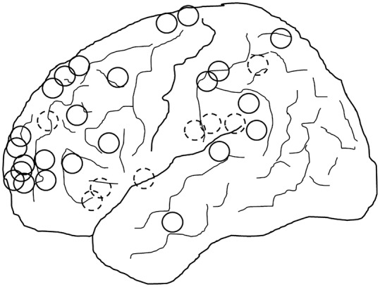
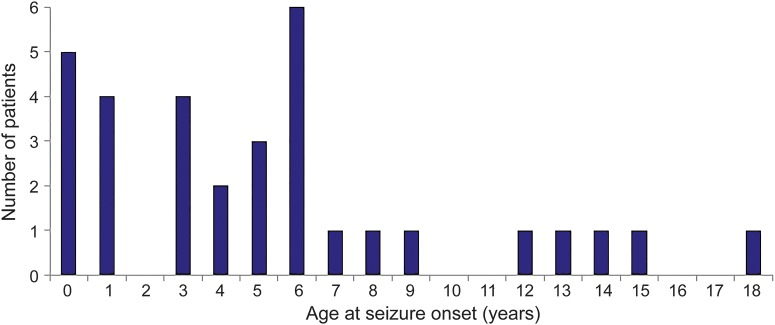
Results: Localization of BOSDs was frontal in 19, insula in 5, parietal in 5, and temporal in 3, on the convexity or interhemispheric surfaces. BOSDs were missed on initial MRI at our centers in 22% of patients. Patients presented with focal seizures during infancy in 9, preschool years in 15, and school years in 8 (median age 5 years). Seizures were stereotyped, predominantly nocturnal, and typically nonconvulsive, with semiology referable to the fronto-central or perisylvian regions. Seizures occurred at high frequency during active periods, but often went into prolonged remission with carbamazepine or phenytoin. Intellect was normal or borderline, except in patients with seizure onset during infancy. Scalp EEG frequently revealed localized interictal epileptiform discharges and ictal rhythms. Patients underwent lesionectomy (median age 14 years) guided by electrocorticography and MRI, with prior intracranial EEG monitoring in only one patient. Twenty-eight patients (88%) became seizure-free, and 20 discontinued antiepileptic medication (median follow-up 4.1 years).

Conclusions: In patients with cryptogenic focal epilepsy, this clinical presentation and course should prompt review of or repeat MRI, looking for a BOSD in the frontal, parietal, or insula cortex. If a BOSD is identified, the patient might be considered for single-stage lesionectomy.


Figures

Figure 1
Figure 1. Three MRI examples of BOSDs
Coronal inversion recovery (A.a) and coronal T2-weighted (A.b) MRI scans at 3.0 tesla (T) showing a BOSD in the right circular sulcus in a 5-year-old girl with asymmetric tonic focal seizures. Coronal FLAIR (B.a) and sagittal FLAIR (B.b) MRI scans at 3.0T showing a BOSD in the right superior parietal lobule in a 17-year-old male with focal motor seizures. Coronal T2-weighted (C.a) and axial FLAIR (C.b) MRI scans at 1.5T showing a BOSD in the left superior frontal gyrus in a 35-year-old man with hypomotor focal seizures. Each example shows cortical thickening, blurred gray–white junction, and subcortical T2/FLAIR hyperintensity at the depth of the sulcus (thick arrow), with a “transmantle sign” extending through the deeper white matter and tapering to the ventricle (thin arrow). Resection of the BOSD identified focal cortical dysplasia type 2B pathology in each case. BOSD = bottom-of-sulcus dysplasia; FLAIR = fluid-attenuated inversion recovery.
Figure 2
Figure 2. Multisequence MRI of a right frontal pole BOSD
Axial MRI scans in the same plane at 3.0 tesla with a 32-channel head coil and T1-weighted fast spoiled gradient recalled echo (A), T2-weighted fast spin echo (B), FLAIR (C), inversion recovery (D), DIR (E), and diffusion-weighted (F) sequences in a 16-year-old boy with hypermotor seizures and right anterior frontal epileptiform activity on scalp EEG. There is a subtle BOSD in the depth of the right superior frontal sulcus anteriorly with thickening of cortex, blurring of gray–white junction, and increased T2/FLAIR/DIR subcortical signal at the bottom of the sulcus (thick arrow) and a “transmantle sign” (thin arrow). Focal cortical dysplasia type 2B pathology was identified at the depth of the resected sulcus. BOSD = bottom-of-sulcus dysplasia; DIR = double inversion recovery; FLAIR = fluid-attenuated inversion recovery.
Figure 3
Figure 3. Coregistered PET and MRI of a right frontal BOSD
Coregistered coronal FDG-PET and T1-weighted MRI scans (A) through the right frontal lobe in a 10-year-old boy with versive seizures and right frontal epileptiform activity on scalp EEG showing localized cortical hypometabolism in the right inferior frontal sulcus. Magnified coronal FLAIR (B) and T2-weighted (C) MRI scans at 3.0 tesla with a 32-channel head coil showing subtle thickening of cortex with blurring of gray–white junction (thick arrow) and faint subcortical signal hyperintensity (hatched arrow) in the bottom of the hypometabolic sulcus, but no “transmantle sign.” The BOSD was not detected on this MRI scan until after coregistration with the PET scan and recognition that there was thickened gray matter deeper than the apparent depth of the hypometabolic sulcus on the PET scan, being more hypometabolic than the sulcal banks. Focal cortical dysplasia type 2B pathology was identified at the depth of the resected sulcus. BOSD = bottom-of-sulcus dysplasia; FDG = fluorodeoxyglucose; FLAIR = fluid-attenuated inversion recovery.
Figure 4
Figure 4. Location of BOSDs
Schematic diagram showing the location of BOSDs in the 32 patients, all represented on the left hemisphere (20 right, 12 left). Full circles represent BOSDs on the cerebral convexity and hatched circles represent BOSDs on the interhemispheric surface and insula. The most common location of BOSDs was in the frontal, parietal, and insula cortex. BOSD = bottom-of-sulcus dysplasia.
Figure 5
Figure 5. Age at seizure onset in 32 patients with BOSD
Age at seizure onset in 32 patients with bottom-of-sulcus dysplasia (BOSD).

Comment in

  • Epilepsy and the funny sulcus.
    Miller JW, Gonzalez-Martinez J. Miller JW, et al. Neurology. 2015 May 19;84(20):2012-3. doi: 10.1212/WNL.0000000000001603. Epub 2015 Apr 17. Neurology. 2015. PMID: 25888555 No abstract available.

References

    1. Barkovich AJ, Kuzniecky RI, Bollen AW, Grant PE. Focal transmantle dysplasia: a specific malformation of cortical development. Neurology 1997;49:1148–1152. - PubMed
    1. Blümcke I, Thom M, Aronica E, et al. The clinicopathologic spectrum of focal cortical dysplasias: a consensus classification proposed by an ad hoc Task Force of the ILAE Diagnostic Methods Commission. Epilepsia 2011;52:158–174. - PMC - PubMed
    1. Barkovich AJ, Guerrini R, Kuzniecky RI, Jackson GD, Dobyns WB. A developmental and genetic classification for malformations of cortical development: update 2012. Brain 2012;135:1348–1369. - PMC - PubMed
    1. Mühlebner A, Coras R, Kobow K, et al. Neuropathologic measurements in focal cortical dysplasias: validation of the ILAE 2011 classification system and diagnostic implications for MRI. Acta Neuropathol 2011;123:259–272. - PubMed
    1. Shepherd C, Liu J, Goc J, et al. A quantitative study of white matter hypomyelination and oligodendroglial maturation in focal cortical dysplasia type II. Epilepsia 2013;54:898–908. - PMC - PubMed

MeSH terms