. 2015 Aug;96(8):2464-2469.
doi: 10.1099/vir.0.000151. Epub 2015 Apr 17.

The placenta shed from goats with classical scrapie is infectious to goat kids and lambs

Affiliations

The placenta shed from goats with classical scrapie is infectious to goat kids and lambs

David A Schneider et al. J Gen Virol. 2015 Aug.

Abstract

The placenta of domestic sheep plays a key role in horizontal transmission of classical scrapie. Domestic goats are frequently raised with sheep and are susceptible to classical scrapie, yet potential routes of transmission from goats to sheep are not fully defined. Sparse accumulation of disease-associated prion protein in cotyledons casts doubt about the role of the goat's placenta. Thus, relevant to mixed-herd management and scrapie-eradication efforts worldwide, we determined if the goat's placenta contains prions orally infectious to goat kids and lambs. A pooled cotyledon homogenate, prepared from the shed placenta of a goat with naturally acquired classical scrapie disease, was used to orally inoculate scrapie-naïve prion genotype-matched goat kids and scrapie-susceptible lambs raised separately in a scrapie-free environment. Transmission was detected in all four goats and in two of four sheep, which importantly identifies the goat's placenta as a risk for horizontal transmission to sheep and other goats.


Figures

Fig. 1.
Fig. 1.. Comparison of the disease-associated prion protein content in the hindbrain at the level of the obex and in shed cotyledons from donor goat 3950. (a) Proteinase K-resistant prion protein (PrPres) bands were detected by Western blot analysis in the obex (ob, lane 1) and cotyledon (cot, lane 2; Na-PTA precipitate) homogenates derived from the scrapie-infected donor goat 3950 but not in similarly prepared PRNP genotype-matched cotyledon homogenate from the scrapie-naïve donor goat 4113 (cot, lane 3; Na-PTA precipitate). (b) Scrapie ELISA titrations of disease-associated prion protein (PrPSc) in shed cotyledons (open diamonds) and obex (open circles) from donor goat 3950. PrPSc content is expressed as background-corrected arbitrary units of optical density (corrected OD); tissue equivalents loaded at each twofold serial dilution are expressed in terms of total protein content (x-axis scaling, log2). As determined per manufacturer's directions, the scrapie ELISA cut-off sensitivity was 0.228 and is shown as a horizontal line.
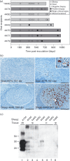
Fig. 2.
Fig. 2.. Detection of classical scrapie transmission in recipient goat kids and lambs. (a) Timeline for recipients orally inoculated with pooled cotyledon homogenate derived from a scrapie-infected goat. Shown are the results of biopsies (circles; open = PrPSc not detected, closed = PrPSc detected), movement from isolation to a quarantine farm environment (#), start of clinical signs of scrapie (solid star) and post-mortem examination (end of bar) of each recipient animal. *Sheep 4440 euthanized due to intercurrent disease (abomasal emptying disorder). (b) Representative images of scrapie IHC (PrPSc = red chromagen deposits) on antemortem biopsies of the rectoanal mucosa-associated lymphoid tissue. Images in which PrPSc was not detected (left column) or was detected (right column) are presented for each recipient species (goat, top row; sheep, bottom row). Age at time of biopsy is given as days post-inoculation (dpi). Accumulation of PrPSc is relatively sparse in goat 4479 as compared with sheep 4442. A higher power inset (bar = 20 μm) of the region of interest (white box) is provided for goat 4479. All other bars = 200 μm. (c) Detection of proteinase K-resistant prion protein (PrPres) bands by Western blot after Na-PTA extraction of retropharyngeal (rp) and ileocecal (ic) lymph node homogenates. The typical three PrPres glycoforms are only evident in the lymph nodes of recipient sheep 4440 (rp, lane 2; ic, lane 5) but not recipient sheep 4445 (lanes 3 and 6) or 4450 (lanes 4 and 7). Assay controls included the Na-PTA extractions of a dilute scrapie-positive obex homogenate (positive control: ob, lane 1) and of a retropharyngeal lymph node from a scrapie uninfected sheep (negative control: rp, lane 8).

References

    1. Alverson J., O'Rourke K.I., Baszler T.V. (2006). PrPSc accumulation in fetal cotyledons of scrapie-resistant lambs is influenced by fetus location in the uterus J Gen Virol 87 1035–1041 10.1099/vir.0.81418-0 . - DOI - PubMed
    1. Andréoletti O., Lacroux C., Chabert A., Monnereau L., Tabouret G., Lantier F., Berthon P., Eychenne F., Lafond-Benestad S., other authors (2002). PrP(Sc) accumulation in placentas of ewes exposed to natural scrapie: influence of foetal PrP genotype and effect on ewe-to-lamb transmission J Gen Virol 83 2607–2616. - PubMed
    1. Baylis M., Chihota C., Stevenson E., Goldmann W., Smith A., Sivam K., Tongue S., Gravenor M.B. (2004). Risk of scrapie in British sheep of different prion protein genotype J Gen Virol 85 2735–2740 10.1099/vir.0.79876-0 . - DOI - PubMed
    1. Blacklaws B.A. (2012). Small ruminant lentiviruses: immunopathogenesis of visna-maedi and caprine arthritis and encephalitis virus Comp Immunol Microbiol Infect Dis 35 259–269 10.1016/j.cimid.2011.12.003 . - DOI - PubMed
    1. Caughey B., Baron G.S., Chesebro B., Jeffrey M. (2009). Getting a grip on prions: oligomers, amyloids, and pathological membrane interactions Annu Rev Biochem 78 177–204 10.1146/annurev.biochem.78.082907.145410 . - DOI - PMC - PubMed

Publication types

LinkOut - more resources