

Tau facilitates Aβ-induced loss of mitochondrial membrane potential independent of cytosolic calcium fluxes in mouse cortical neurons
- PMID: 25888814
- PMCID: PMC4442715
- DOI: 10.1016/j.neulet.2015.04.021
Tau facilitates Aβ-induced loss of mitochondrial membrane potential independent of cytosolic calcium fluxes in mouse cortical neurons
Abstract
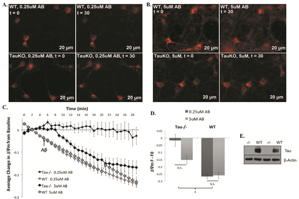
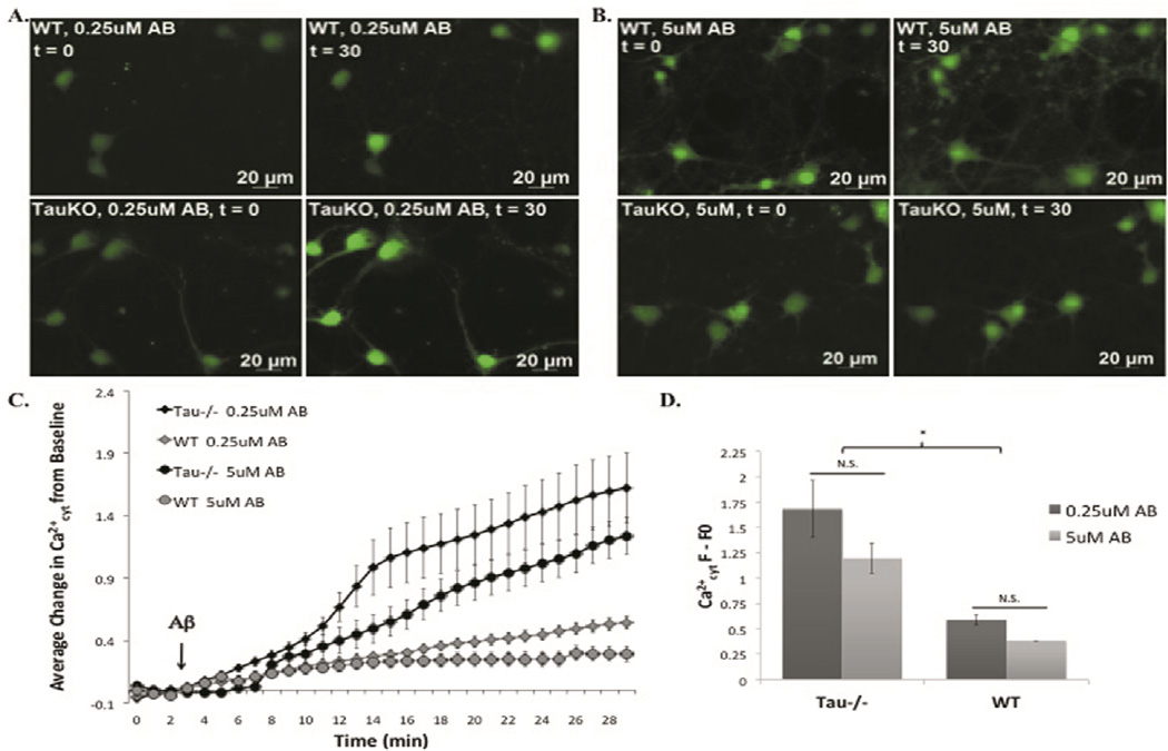
Alzheimer's disease (AD) is defined by presence of two pathological hallmarks, the intraneuronal neurofibrillary tangle (NFT) formed by abnormally processed tau, and the extracellular amyloid plaques formed primarily by the amyloid beta peptide (Aβ). In AD it is likely that these two proteins act in concert to impair neuronal function, and there is evidence to suggest that one of the key targets on which they converge is the mitochondria. For example, overexpression of a pathologic form of tau in rat primary cortical neurons exacerbates Aβ-induced mitochondrial membrane potential (ΔΨm) loss due to impairment of the calcium (Ca(2+)) buffering capability of mitochondria. However the role of physiological levels of tau in mediating Aβ-induced mitochondrial dysfunction was not examined. Therefore in this present study we used primary neurons from wild type (WT) and tau knockout (tau(-/-)) mice to investigate whether endogenous tau facilitates Aβ-induced ΔΨm loss and alterations in cytosolic calcium (Ca(2+)cyt). Knocking out tau significantly protected mouse primary cortical neurons from loss of ΔΨm caused by low concentrations of Aβ42, which supports our previous findings. However, the absence of tau resulted in significantly greater increases in Ca(2+)cyt in response to Aβ treatment when compared to those observed in WT mouse primary cortical neurons. This unexpected outcome may be explained by findings that suggest tau(-/-) neurons display certain phenotypic abnormalities associated with alterations in Ca(2+)cyt. Overall, data indicate that tau facilitates Aβ-induced mitochondrial dysfunction and this effect is independent of Aβ-induced alterations in Ca(2+)cyt.(1).
Keywords: Alzheimer’s disease; Amyloid beta; Calcium dyshomeostasis; Mitochondrial dysfunction; Tau.
Copyright © 2015 Elsevier Ireland Ltd. All rights reserved.
Figures
References
-
- Bubber P, Haroutunian V, Fisch G, Blass JP, Gibson GE. Mitochondrial abnormalities in Alzheimer brain: mechanistic implications. Ann Neurol. 2005;57:695–703. - PubMed
-
- Davis JN, Hunnicutt EJ, Jr, Chisholm JC. A mitochondrial bottleneck hypothesis of Alzheimer's disease. Molecular medicine today. 1995;1:240–247. - PubMed
-
- Dawson HN, Ferreira A, Eyster MV, Ghoshal N, Binder LI, Vitek MP. Inhibition of neuronal maturation in primary hippocampal neurons from tau deficient mice. J Cell Sci. 2001;114:1179–1187. - PubMed
Publication types
MeSH terms
Substances
Grants and funding
LinkOut - more resources
Full Text Sources
Other Literature Sources
Miscellaneous
