. 2015 Mar 6:46:26.
doi: 10.1186/s13567-015-0154-7.

Piscine orthoreovirus (PRV) replicates in Atlantic salmon (Salmo salar L.) erythrocytes ex vivo

Affiliations

Piscine orthoreovirus (PRV) replicates in Atlantic salmon (Salmo salar L.) erythrocytes ex vivo

Øystein Wessel et al. Vet Res. .

Abstract

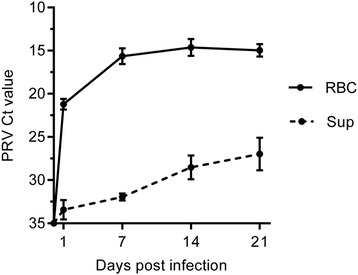
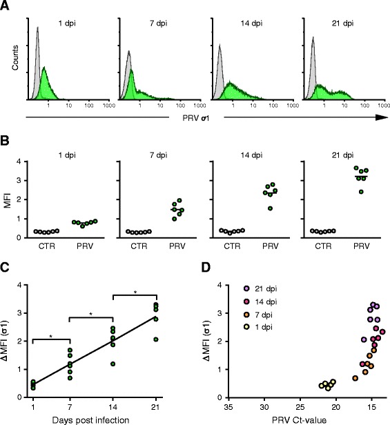
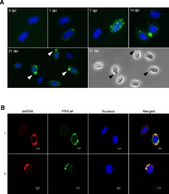
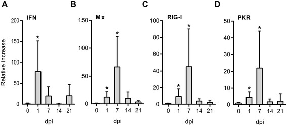
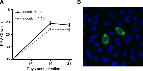
Piscine orthoreovirus (PRV) is a reovirus that has predominantly been detected in Atlantic salmon (Salmo salar L.). PRV is associated with heart and skeletal muscle inflammation (HSMI) in farmed Atlantic salmon, and recently erythrocytes were identified as major target cells. The study of PRV replication and pathogenesis of the infection has been impeded by the inability to propagate PRV in vitro. In this study we developed an ex vivo cultivation system for PRV in Atlantic salmon erythrocytes. PRV was successfully passaged to naïve erythrocytes using lysates of blood cells from infected salmon. During cultivation a significant increase in viral load was observed by RT-qPCR and flow cytometry, which coincided with the formation of cytoplasmic inclusions. The inclusions resembled viral factories and contained both PRV protein and dsRNA. In addition, the erythrocytes generated an antiviral immune gene activation after PRV infection, with significant up-regulation of IFN-α, RIG-I, Mx and PKR transcripts. Supernatants from the first passage successfully transmitted virus to naïve erythrocytes. This study demonstrates that PRV replicates in Atlantic salmon erythrocytes ex vivo. The ex vivo infection model closely reflects the situation in vivo and can be used to study the infection and replication mechanisms of PRV, as well as the antiviral immune responses of salmonid erythrocytes.


Figures

Figure 1
Figure 1
PRV load in 1 st passage of culturing. Mean PRV Ct value with SD (n = 6) as detected by RT-qPCR in red blood cells (RBC) and supernatant (Sup) at 1, 7, 14 and 21 days post infection.
Figure 2
Figure 2
Detection of PRV ơ1 protein by flow cytometry in the 1 st passage. (A) Results from intracellular staining for PRV ơ1 protein from PRV infected culture (green) and control culture (grey) at 1, 7, 14 and 21 days post infection (dpi). The fluorescence intensity (PRV ơ1) is shown on the X-axis and cell count on the y-axis counting 30 000 cells per sample. The results of one individual are presented to illustrate the staining pattern. (B) Plot of the arithmetic mean fluorescence intensity (MFI) for all PRV infected (green) and control (grey) cultures (n = 6) at each time point. (C) The amount of PRV ơ1 protein detected presented by ΔMFI (MFI Infected – MFI Control) at 1, 7, 14 and 21 dpi. Data were analyzed using Wilcoxon matched pairs signed rank test. *p < 0.05. (D) The correlation between the amount of PRV nucleic acid and PRV ơ1 protein detected during the cultivation is illustrated by plotting PRV Ct-values (X-axis) against the ΔMFI for PRV ơ1 (Y-axis) color coding the different time points; 1 dpi (yellow), 7 dpi (orange), 14 dpi (red) and 21 dpi (purple).
Figure 3
Figure 3
Immunofluorescence and confocal microscopy of PRV infected RBC. (A) Fluorescent labeling of the PRV ơ1-protein (green) in ex vivo infected RBC. The nuclei were stained with Hoechst (blue). Non-infected cells are shown at 0 dpi, and infected cells are shown at 1, 7 14 and 21 dpi. Large PRV inclusions at 21 dpi (marked by arrowheads) can also be observed in phase contrast. (B) Confocal images of ex vivo infected RBC at 21 dpi, stained with anti-dsRNA (red) and anti-PRV-ơ1 (green). The nuclei were stained with Hoechst (blue). dsRNA was detected within PRV inclusions, and either partly (i) or completely (ii) co-localized with PRV ơ1. Scale bar 2 μm.
Figure 4
Figure 4
Erythrocytic antiviral responses to PRV infection. Expression of genes involved in antiviral responses was measured by RT-qPCR. The expression levels in infected RBC relative to the paired non-infected controls were calculated for each sample (n = 6) at 1, 7, 12 and 21 dpi. The relative increase (and SD) for IFNα (A), Mx (B), RIG-I (C) and PKR (D) is shown. Data were analyzed using Wilcoxon matched pairs signed rank test. *p < 0.05.
Figure 5
Figure 5
RT-qPCR and PRV ơ1 staining from the 2 nd passage. (A) Mean PRV Ct value (and SD) detected by RT-qPCR in red blood cells (RBC) at 14 and 21 dpi (n = 6) during the second passage. Results are shown for cultures infected with undiluted and diluted (1:10) inoculum from the 1st passage. (B) Immunofluorescence microscopy picture showing the PRV ơ1-protein (green) in cells at 14 dpi infected by undiluted inoculum. Cell nuclei stained with Hoechst (blue).

References

    1. Palacios G, Løvoll M, Tengs T, Hornig M, Hutchison S, Hui J, Kongtorp RT, Savji N, Bussetti AV, Solovyov A, Kristoffersen AB, Celone C, Street C, Trifonov V, Hirschberg DL, Rabadan R, Egholm M, Rimstad E, Lipkin WI. Heart and skeletal muscle inflammation of farmed salmon is associated with infection with a novel reovirus. PLoS One. 2010;5:e11487. doi: 10.1371/journal.pone.0011487. - DOI - PMC - PubMed
    1. Kongtorp RT, Kjerstad A, Taksdal T, Guttvik A, Falk K. Heart and skeletal muscle inflammation in Atlantic salmon, Salmo salar L.: a new infectious disease. J Fish Dis. 2004;27:351–358. doi: 10.1111/j.1365-2761.2004.00549.x. - DOI - PubMed
    1. Løvoll M, Alarcon M, Bang JB, Taksdal T, Kristoffersen AB, Tengs T. Quantification of piscine reovirus (PRV) at different stages of Atlantic salmon Salmo salar production. Dis Aquat Organ. 2012;99:7–12. doi: 10.3354/dao02451. - DOI - PubMed
    1. Kibenge MJ, Iwamoto T, Wang Y, Morton A, Godoy MG, Kibenge FS. Whole-genome analysis of piscine reovirus (PRV) shows PRV represents a new genus in family Reoviridae and its genome segment S1 sequences group it into two separate sub-genotypes. Virol J. 2013;10:230. doi: 10.1186/1743-422X-10-230. - DOI - PMC - PubMed
    1. Marty GD, Morrison DB, Bidulka J, Joseph T, Siah A. Piscine reovirus in wild and farmed salmonids in British Columbia, Canada: 1974–2013. J Fish Dis, in press. - PubMed

LinkOut - more resources