Candesartan restores pressure-induced vasodilation and prevents skin pressure ulcer formation in diabetic mice
- PMID: 25888905
- PMCID: PMC4394592
- DOI: 10.1186/s12933-015-0185-4
Candesartan restores pressure-induced vasodilation and prevents skin pressure ulcer formation in diabetic mice
Abstract
Background: Angiotensin II type 1 receptor (AT1R) blockers have beneficial effects on neurovascular complications in diabetes and in organ's protection against ischemic episodes. The present study examines whether the AT1R blocker candesartan (1) has a beneficial effect on diabetes-induced alteration of pressure-induced vasodilation (PIV, a cutaneous physiological neurovascular mechanism which could delay the occurrence of tissue ischemia), and (2) could be protective against skin pressure ulcer formation.
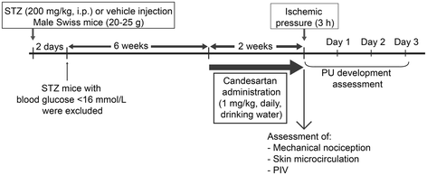
Methods: Male Swiss mice aged 5-6 weeks were randomly assigned to four experimental groups. In two groups, diabetes was induced by a single intraperitoneal injection of streptozotocin (STZ, 200 mg.kg(-1)). After 6 weeks, control and STZ mice received either no treatment or candesartan (1 mg/kg-daily in drinking water) during 2 weeks. At the end of treatment (8 weeks of diabetes duration), C-fiber mediated nociception threshold, endothelium-dependent vasodilation and PIV were assessed. Pressure ulcers (PUs) were then induced by pinching the dorsal skin between two magnetic plates for three hours. Skin ulcer area development was assessed during three days, and histological examination of the depth of the skin lesion was performed at day three.
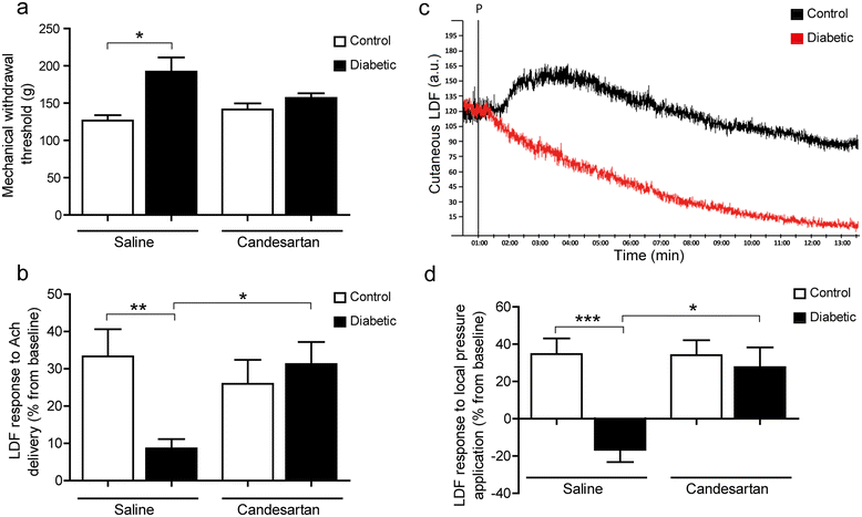
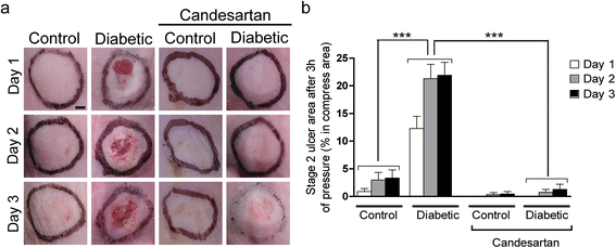
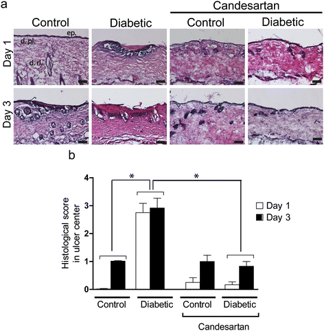
Results: After 8 weeks of diabetes, the skin neurovascular functions (C-fiber nociception, endothelium-dependent vasodilation and PIV) were markedly altered in STZ-treated mice, but were fully restored by treatment with candesartan. Whereas in diabetes mice exposure of the skin to pressure induced wide and deep necrotic lesions, treatment with candersartan restored their ability to resist to pressure-induced ulceration as efficiently as the control mice.
Conclusion: Candesartan decreases the vulnerability to pressure-induced ulceration and restores skin neurovascular functions in mice with STZ-induced established diabetes.
Figures


Similar articles
-
Erythropoietin restores C-fiber function and prevents pressure ulcer formation in diabetic mice.J Invest Dermatol. 2011 Nov;131(11):2316-22. doi: 10.1038/jid.2011.211. Epub 2011 Aug 11. J Invest Dermatol. 2011. PMID: 21833012
-
Protective mechanisms of the angiotensin II type 1 receptor blocker candesartan against cerebral ischemia: in-vivo and in-vitro studies.J Hypertens. 2008 Jul;26(7):1435-45. doi: 10.1097/HJH.0b013e3283013b6e. J Hypertens. 2008. PMID: 18551021
-
Angiotensin II type 1 receptor blocker attenuates diabetes-induced atrial structural remodeling.J Cardiol. 2011 Sep;58(2):131-6. doi: 10.1016/j.jjcc.2011.06.003. Epub 2011 Jul 30. J Cardiol. 2011. PMID: 21802905
-
Candesartan: widening indications for this angiotensin II receptor blocker?Expert Opin Pharmacother. 2009 Aug;10(12):1995-2007. doi: 10.1517/14656560903092197. Expert Opin Pharmacother. 2009. PMID: 19563275 Review.
-
Newborn and elderly skin: two fragile skins at higher risk of pressure injury.Biol Rev Camb Philos Soc. 2022 Jun;97(3):874-895. doi: 10.1111/brv.12827. Epub 2021 Dec 15. Biol Rev Camb Philos Soc. 2022. PMID: 34913582 Review.
Cited by
-
Vaccarin Regulates Diabetic Chronic Wound Healing through FOXP2/AGGF1 Pathways.Int J Mol Sci. 2020 Mar 13;21(6):1966. doi: 10.3390/ijms21061966. Int J Mol Sci. 2020. PMID: 32183046 Free PMC article.
-
[Research progress on the animal models and treatment strategies of diabetic foot ulcer].Zhejiang Da Xue Xue Bao Yi Xue Ban. 2017 Jan 25;46(1):97-105. doi: 10.3785/j.issn.1008-9292.2017.02.15. Zhejiang Da Xue Xue Bao Yi Xue Ban. 2017. PMID: 28436638 Free PMC article. Review. Chinese.
-
Observational case-control study of small-fiber neuropathies, with regards on smoking and vitamin D deficiency and other possible causes.Front Med (Lausanne). 2023 Jan 12;9:1051967. doi: 10.3389/fmed.2022.1051967. eCollection 2022. Front Med (Lausanne). 2023. PMID: 36714112 Free PMC article.
-
Pressure-induced vasodilation mimicking vasculopathy in a pediatric patient.JAAD Case Rep. 2016 Feb 27;2(1):87-8. doi: 10.1016/j.jdcr.2015.12.004. eCollection 2016 Jan. JAAD Case Rep. 2016. PMID: 27051838 Free PMC article. No abstract available.
-
Scorpion Venom Active Polypeptide May Be a New External Drug of Diabetic Ulcer.Evid Based Complement Alternat Med. 2017;2017:5161565. doi: 10.1155/2017/5161565. Epub 2017 Oct 29. Evid Based Complement Alternat Med. 2017. PMID: 29234410 Free PMC article.
References
Publication types
MeSH terms
Substances
LinkOut - more resources
Full Text Sources
Other Literature Sources
Medical

