. 2015 Mar 24;6(1):44.
doi: 10.1186/s13287-015-0025-1.

Interaction between mesenchymal stem cells and endothelial cells restores endothelial permeability via paracrine hepatocyte growth factor in vitro

Affiliations

Interaction between mesenchymal stem cells and endothelial cells restores endothelial permeability via paracrine hepatocyte growth factor in vitro

Qi-Hong Chen et al. Stem Cell Res Ther. .

Abstract

Introduction: Mesenchymal stem cells (MSCs) have potent stabilising effects on vascular endothelium injury, inhibiting endothelial permeability in lung injury via paracrine hepatocyte growth factor (HGF). Recently, it has been indicated that MSCs secrete more factors by MSC-endothelial cell (MSC-EC) interactions. We hypothesised that MSC-EC interactions restore endothelial permeability induced by lipopolysaccharide (LPS) via paracrine HGF.

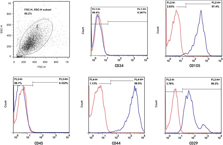
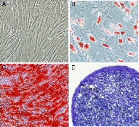
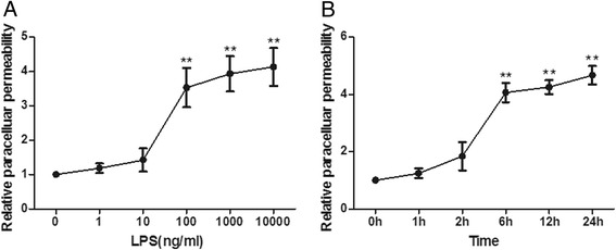
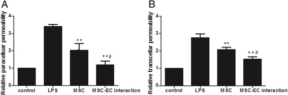
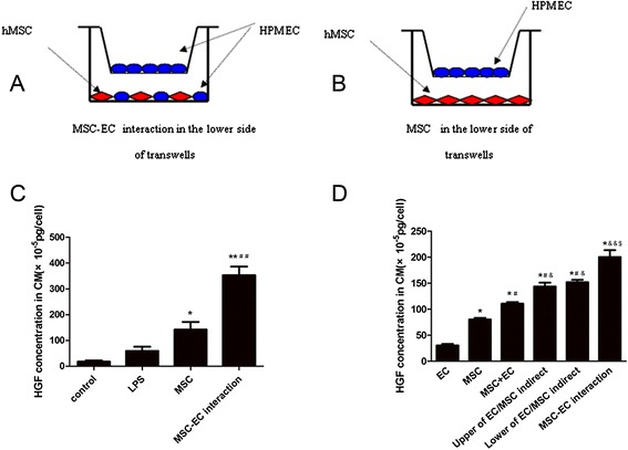
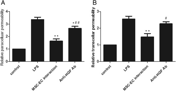
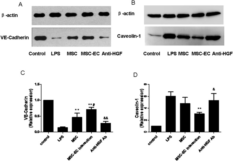
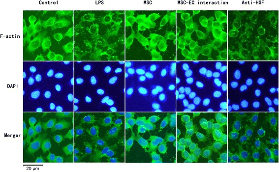
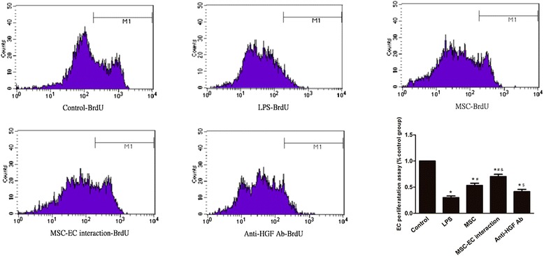
Methods: We investigated the endothelial permeability induced by LPS under two co-culture conditions. Human pulmonary microvascular endothelial cells (HPMECs) were added into the upper chambers of cell-culture inserts, while two different co-culture conditions were used in the lower side of the transwells, as follows: (1) MSC-EC interaction group: MSCs and HPMECs contact co-culture; (2) MSC group: MSCs only. The endothelial paracellular and transcellular permeabilities in the upper side of transwells were detected. Then the concentration of HGF was measured in the culture medium by using an enzyme-linked immunosorbent assay kit, followed by neutralisation of HGF with anti-HGF antibody in the co-culture medium. In addition, adherens junction and cytoskeleton protein expressions were measured by Western blot and immunofluorescence. HPMEC proliferation was analysed by bromodeoxyuridine incorporation assay.

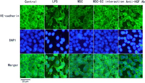
Results: The paracellular permeability significantly increased after LPS stimulation in a dose-dependent and time-dependent manner. Meanwhile, MSC-EC interaction more significantly decreased endothelial paracellular and transcellular permeability induced by LPS. Moreover, HGF levels in the MSC-EC interaction group were much higher than those of the MSC group. However, neutralising HGF with anti-HGF antibody inhibited the role of MSC-EC interaction in improving endothelial permeability. Compared with the MSC group, MSC-EC interaction increased vascular endothelial (VE)-cadherin and occludin protein expression, reduced caveolin-1 protein expression in HPMECs, and restored remodelling of F-actin and junctional localisation of VE-cadherin. Furthermore, the proliferation ratio in the MSC-EC interaction group was higher than that of the MSC group. However, the effects of MSCs were significantly blocked by anti-HGF antibody.

Conclusions: These data suggested that MSC-EC interaction decreased endothelial permeability induced by LPS, which was attributed mainly to HGF secreted by MSCs. The main mechanisms by which HGF restored the integrity of endothelial monolayers were remodelling of endothelial intercellular junctions, decreasing caveolin-1 protein expression, and inducing proliferation in HPMECs.


Figures

Figure 1
Figure 1
Flow cytometry identification of human mesenchymal stem cells (hMSCs). Cell surface markers of hMSCs, including CD34, CD44, CD105, CD29, and CD45, were analysed with flow cytometry. Red lines represent the isotype controls. The supplier guarantees that there is no ethical issue in using the hMSCs for experiments. FSC-H, forward scatter height; SSC-H, side scatter height.
Figure 2
Figure 2
Multilineage differentiation identification of human mesenchymal stem cells. Morphology of human mesenchymal stem cells at the third passage (A) (100 T) and multilineage differentiation capacities of mouse mesenchymal stem cells, including adipogenic differentiation stained with Oil Red O (B) (200 T), osteogenic differentiation stained with Alizarin red (C) (200 T), and chondrogenic differentiation stained with Toluidine blue (D) (200 T), were observed by microscope.
Figure 3
Figure 3
Lipopolysaccharide (LPS) evokes human pulmonary microvascular endothelial cell hyperpermeability. The paracellular permeability of human pulmonary microvascular endothelial cells was measured at different LPS dosages and at different time points after LPS intervention. The results showed that paracellular permeability had no significant alterations after LPS addition with dosages below 10 ng/mL. However, the paracellular permeability significantly increased after LPS stimulation with dosages above 100 ng/mL in a dose-dependent manner (A). Furthermore, we observed that paracellular permeability significantly increased after 6 hours following 100 ng/mL LPS addition in a time-dependent manner (B). Shown are the mean ± standard error of three parallel experiments (n = 3, **P <0.01 versus group 0 ng/mL or 0 hours).
Figure 4
Figure 4
Mesenchymal stem cell-endothelial cell (MSC-EC) interaction promoted human MSCs to restore the integrity of human pulmonary microvascular endothelial cell (HPMEC) monolayers. We found HPMEC paracellular permeability for both MSC-EC interaction and MSC groups significantly decreased (P <0.01) (A). In addition, HPMEC paracellular permeability in the MSC-EC interaction group decreased more significantly than that of the MSC group (P <0.05) (A). Similarly, HPMEC transcellular permeability significantly decreased in two co-culture systems (P <0.01) (B), while MSC-EC interaction more significantly decreased HPMEC transcellular permeability (P <0.05) (B). (n=3, ** p < 0.01 vs. group LPS; # p < 0.05 vs. group MSC). LPS, lipopolysaccharide.
Figure 5
Figure 5
More hepatocyte growth factor (HGF) was secreted under mesenchymal stem cell-endothelial cell (MSC-EC) interactions. We adopted two different co-culture conditions through transwell insert including MSC-EC interactions (A) and MSCs (B). Then we measured the concentration of HGF in the culture medium by using an enzyme-linked immunosorbent assay (ELISA) kit. The results indicated that the concentration of HGF in direct co-culture and indirect co-culture media was significantly higher than that of the lipopolysaccharide (LPS) group. Interestingly, HGF levels in direct co-culture medium were much higher than that of indirect co-culture medium (C) (n = 3, **P <0.01 versus LPS group; ## P <0.01 versus indirect co-culture group). We further tested HGF concentration by ELISA quantification in conditioned medium from six different formats. The results showed that the concentration of HGF in the MSC group was significantly higher than that of the EC group. HGF levels had no significant difference between the upper and lower of the MSC-EC indirect co-culture group, which were higher compared with the MSC group and the sum of the human pulmonary microvascular endothelial cell (HPMEC) and MSC alone group (D) (P <0.05 and P <0.01). However, HGF levels in the MSC-EC interaction group were highest in all six groups (D) (P <0.01) (n = 3, *P <0.01 versus EC group, # P <0.01 versus MSC group; & P <0.05, && P <0.01 versus MSC + EC group, $ P <0.01 versus upper of MSC-EC indirect group or lower of MSC-EC indirect group). CM, conditioned media; hMSC, human mesenchymal stem cell.
Figure 6
Figure 6
Hepatocyte growth factor (HGF) neutralisation attenuated the restoration of the integrity of human pulmonary microvascular endothelial cell (HPMEC) monolayers in mesenchymal stem cell-endothelial cell (MSC-EC) interaction conditions. We found that the paracellular and transcellular permeability in the anti-HGF antibody (Ab) group was significantly increased over control (without anti-HGF Ab) (n = 3, P <0.05) (A, B), suggesting that human MSCs restored the integrity of HPMEC monolayers in MSC-EC interaction by paracrine HGF. (n=3, ** p < 0.01 vs. group LPS; # p<0.05 # # p < 0.01 vs. group MSC-EC interaction) LPS, lipopolysaccharide.
Figure 7
Figure 7
Hepatocyte growth factor (HGF) secreted by human mesenchymal stem cells (hMSCs) following mesenchymal stem cell-endothelial cell (MSC-EC) interactions upregulated vascular endothelial (VE)-cadherin protein and decreased caveolin-1 protein expression. Compared with the MSC group, MSC-EC interaction increased VE-cadherin protein expression (P <0.01) (A, C) while reducing caveolin-1 protein expression in human pulmonary microvascular endothelial cells (HPMECs) (P <0.01) (B, D). However, the MSC effect was significantly blocked by anti-HGF antibody (P <0.05 or P <0.01) (C, D). These results indicated that HGF secreted by hMSCs upregulated endothelial VE-cadherin protein and decreased caveolin-1 protein expression. (n=3, * *p < 0.01 vs. group LPS, # p < 0.01 vs. MSC; & p< 0.05 && p < 0.01 vs. group MSC-EC interaction) LPS, lipopolysaccharide.
Figure 8
Figure 8
Hepatocyte growth factor (HGF) secreted in mesenchymal stem cell-endothelial cell (MSC-EC) interactions conditions restored remodelling of endothelial F-actin. The results indicated that human pulmonary microvascular endothelial cells cultured under normal condition had F-actin localised mainly at the cell periphery. Lipopolysaccharide (LPS) induced dispersion of F-actin. After 7 days of direct or indirect co-culture with human MSCs, remodelling of F-actin was partially restored. However, neutralising HGF from conditioned media with anti-HGF antibody caused F-actin to be disrupted again. DAPI, 4,6-diamidino-2-phenylindole.
Figure 9
Figure 9
Hepatocyte growth factor (HGF) was secreted in mesenchymal stem cell-endothelial cell (MSC-EC) interaction conditions and restored remodelling of vascular endothelial (VE)-cadherin. The results indicated that human pulmonary microvascular endothelial cells cultured under normal conditions had VE-cadherin, the main component of adherens junctions, forming intercellular junctions. Lipopolysaccharide (LPS) induced dispersion of VE-cadherin. After 7 days of direct or indirect co-cultured with human MSCs, VE-cadherin was partially restored. However, neutralising HGF from conditioned media with anti-HGF antibody (Ab) caused VE-cadherin to be disrupted again. DAPI, 4,6-diamidino-2-phenylindole.
Figure 10
Figure 10
Hepatocyte growth factor (HGF) secreted by mesenchymal stem cell-endothelial cell (MSC-EC) interaction induces proliferation in human pulmonary microvascular endothelial cells (HPMECs). To explicit effect of HGF secreted by MSCs on HPMEC proliferation, cell proliferation was analysed by bromodeoxyuridine (BrdU) incorporation assay. The results indicated that MSCs induce proliferation in HPMECs injured by lipopolysaccharide (LPS). Interestingly, the proliferation ratio in the MSC-EC interaction group was higher than that of the MSC group (P <0.05). However, the MSC-EC interaction effect was significantly blocked by anti-HGF antibody (P <0.01), which explains why there are more HPMECs in the MSC-EC group. The result suggested that HGF secreted by MSC-EC interaction induces proliferation in HPMECs (n = 3, *P <0.01 versus control group, # P <0.01 versus LPS group; & P <0.05 versus MSC group, $ P <0.01 versus MSC-EC interaction).

Similar articles

Cited by

References

    1. Gonzales JN, Gorshkov B, Varn MN, Zemskova MA, Zemskov EA, Sridhar S, et al. Protective effect of adenosine receptors against lipopolysaccharide-induced acute lung injury. Am J Physiol Lung Cell Mol Physiol. 2014;306:L497–507. doi: 10.1152/ajplung.00086.2013. - DOI - PMC - PubMed
    1. Pittet JF, Mackersie RC, Martin TR, Matthay MA. Biological markers of acute lung injury: prognostic and pathogenetic significance. Am J Respir Crit Care Med. 1997;155:1187–205. doi: 10.1164/ajrccm.155.4.9105054. - DOI - PubMed
    1. Mei SH, McCarter SD, Deng Y, Parker CH, Liles WC, Stewart DJ. Prevention of LPS-induced acute lung injury in mice by mesenchymal stem cells overexpressing angiopoietin 1. PLoS Med. 2007;4:e269. doi: 10.1371/journal.pmed.0040269. - DOI - PMC - PubMed
    1. Lee JW, Fang X, Gupta N, Serikov V, Matthay MA. Allogeneic human mesenchymal stem cells for treatment of E. coli endotoxin-induced acute lung injury in the ex vivo perfused human lung. Proc Natl Acad Sci U S A. 2009;106:16357–62. doi: 10.1073/pnas.0907996106. - DOI - PMC - PubMed
    1. Song L, Xu J, Qu J, Sai Y, Chen C, Yu L, et al. A therapeutic role for mesenchymal stem cells in acute lung injury independent of hypoxia-induced mitogenic factor. J Cell Mol Med. 2012;16:376–85. doi: 10.1111/j.1582-4934.2011.01326.x. - DOI - PMC - PubMed

Publication types

MeSH terms