. 2015 Mar 28:12:29.
doi: 10.1186/s12977-015-0154-8.

Balanced splicing at the Tat-specific HIV-1 3'ss A3 is critical for HIV-1 replication

Affiliations

Balanced splicing at the Tat-specific HIV-1 3'ss A3 is critical for HIV-1 replication

Steffen Erkelenz et al. Retrovirology. .

Abstract

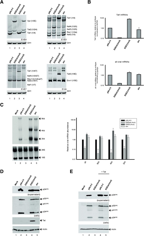
Background: The viral regulatory protein Tat is essential for establishing a productive transcription from the 5'-LTR promoter during the early phase of viral gene expression. Formation of the Tat-encoding mRNAs requires splicing at the viral 3'ss A3, which has previously been shown to be both negatively and positively regulated by the downstream splicing regulatory elements (SREs) ESS2p and ESE2/ESS2. However, using the novel RESCUE-type computational HEXplorer algorithm, we were recently able to identify another splicing enhancer (ESE(5807-5838), henceforth referred to as ESE tat ) located between ESS2p and ESE2/ESS2. Here we show that ESE tat has a great impact on viral tat-mRNA splicing and that it is fundamental for regulated 3'ss A3 usage.

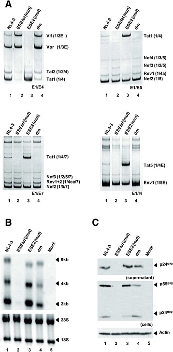
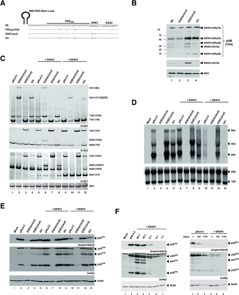
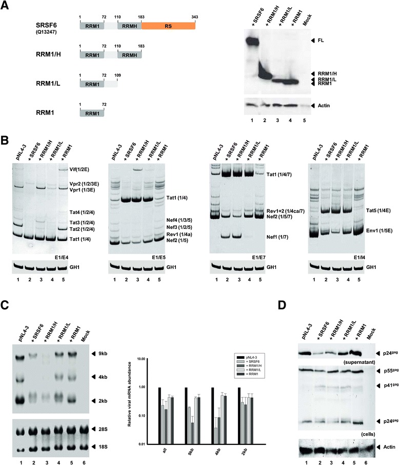
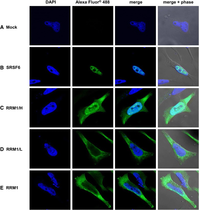
Results: Mutational inactivation or locked nucleic acid (LNA)-directed masking of the ESE tat sequence in the context of a replication-competent virus was associated with a failure (i) to activate viral 3'ss A3 and (ii) to accumulate Tat-encoding mRNA species. Consequently, due to insufficient amounts of Tat protein efficient viral replication was drastically impaired. RNA in vitro binding assays revealed SRSF2 and SRSF6 as candidate splicing factors acting through ESE tat and ESE2 for 3'ss A3 activation. This notion was supported by coexpression experiments, in which wild-type, but not ESE tat -negative provirus responded to higher levels of SRSF2 and SRSF6 proteins with higher levels of tat-mRNA splicing. Remarkably, we could also find that SRSF6 overexpression established an antiviral state within provirus-transfected cells, efficiently blocking virus particle production. For the anti-HIV-1 activity the arginine-serine (RS)-rich domain of the splicing factor was dispensable.

Conclusions: Based on our results, we propose that splicing at 3'ss A3 is dependent on binding of the enhancing SR proteins SRSF2 and SRSF6 to the ESE tat and ESE2 sequence. Mutational inactivation or interference specifically with ESE tat activity by LNA-directed masking seem to account for an early stage defect in viral gene expression, probably by cutting off the supply line of Tat that HIV needs to efficiently transcribe its genome.


Figures

Figure 1
Figure 1
Splicing regulatory elements (SREs) in the HIV-1 genome and mutational analysis of ESE tat . (A) Top: The open reading frames (ORFs) are indicated by open boxes. The long terminal repeats (LTR) are located at both ends of the provirus. Center: All HIV-1 proteins are encoded in a single primary transcript. More than 40 different viral mRNAs are produced by alternative splicing allowing efficient translation of all ORFs within the infected cell. Intrinsic strength of the 5′ss (D1 to D4) and 3′ss (A1-A7) is indicated in parentheses (5′ss: HBond Score, http://www.uni-duesseldorf.de/rna; 3′ss: MaxEntScore, http://genes.mit.edu/burgelab/maxent/Xmaxentscan_scoreseq_acc.html). Bottom: Positions of the SREs within the HIV-1 pre-mRNA: splicing enhancers (green) and silencers (red) are indicated. [ESE705-29 [14,40]; ESE-Vif [41]; ESEM [39]; ESE4932-62 [14]; ESE5005-32 [14]; guanosine (G) rich silencer G4 [41]; GI2-1 [42]; ESSV [43-45]; ESEvpr [12]; GI3-2 [16]; ESS2p [46]; ESE2 [15,47]; ESS2 [48-50]; guanosine-adenosine rich (GAR) ESE [13,37,51]; E42 fragment [51]; ISS [52]; ESE3 [53]; ESS3 [53-55] (adapted to [51,56]). Primers used in RT-PCR analyses are indicated by arrows (forward: E1, reverse: E4, E5, I4 and E7). (B) Top: HIV-1 exon 4 reference (pNL4-3) and mutant sequences used in this study. The ESEtat is indicated by a grey rectangle. Previously published SREs in this region are underlined. Bottom: HEXplorer score profiles for wild-type HIV-1 exon 4 reference (white) and mutant sequences (black). ESEtat is indicated by a grey rectangle. ESE2 is indicated by curly braces. Positions of mutated nucleotides are indicated by arrows.
Figure 2
Figure 2
ESE tat is required for activation of Tat-specific 3′ss A3. (A) 2.5 × 10⁵ HEK293T cells were transiently transfected with each of the proviral plasmids. 48 h post transfection, total RNA was isolated from the cells and analyzed by RT-PCR using different sets of primer pairs (primer positions are shown in Figure 1A). HIV-1 mRNA species are indicated to the right of the gel images according to the nomenclature published previously [6]. (B) Real-time PCR assays to specifically quantitate the relative levels of tat-mRNAs (top) and all viral mRNAs (bottom). For normalization we monitored the total amount of cellular GAPDH present in every sample. Data represent expression ratios relative to that of wild-type pNL4-3 (bar 1). Values and error bars show the average ± standard deviation of three independent transfection experiments. (C) Left: Northern blot analysis of total RNA isolated from the same RNA preparation as in (A). A hybridization probe was used specifically detecting HIV-1 exon 7. Right: Quantification of Northern blot using RNAs from three independently performed transfection experiments. Data represent expression ratios relative to that of wild-type pNL4-3 (bar 1), which was set to 1. For normalization the ribosomal RNA amount of each sample was calculated. Values and error bars show the average ± standard deviation of three independent transfection experiments. (D) Western blot analysis of viral Gag and Tat expressed by wild-type reference and mutant provirus. Supernatants and lysates were probed with a primary antibody against HIV-1 p24gag or HIV-1 tat. Equal amounts of cell lysates were controlled by the detection of α-actin. (E) 2.5 × 10⁵ HEK293T cells were transfected with 1 μg of proviral plasmids and 0.5 μg of pcDNA3.1(+) or SVctat expressing viral Tat protein from a cDNA. Western blot analysis was performed as described in (D).
Figure 3
Figure 3
ESE tat -negative virus fails to efficiently replicate in T-cells. (A) RT-PCR analysis of viral mRNAs taken from Jurkat T-cells that were infected with 10 ng p24gag of wild-type or mutant NL4-3 virus. Total RNA was isolated 6 days post infection and subjected to RT-PCR analysis using different sets of primer pairs (primer positions are shown in Figure 1A). (B) Northern blot analysis of RNAs isolated in (A) using a DIG-labeled HIV-1 exon7 probe detecting all three viral mRNA species. (C) Western blot analysis of intracellular and supernatant HIV-1 Gag collected 6 days post infection as described above. Actin detection was used as loading control.
Figure 4
Figure 4
LNA-mediated masking of ESE tat mimics the influence of the mutated ESE tat on the viral splicing pattern and viral particle production. (A) Schematic illustration of the location of ESS2p, ESEtat, ESE2 and ESS2 as well as the binding site for the locked nucleic acids (LNAs) directed against ESEtat and ESE2/ESS2 sequences. (B) RT-PCR analysis of viral mRNA classes. HeLa cells were transiently transfected with pNL4-3 and either ESEtat-LNA, the ESE2/ESS2 LNA or a scrambled LNA. Total RNA was isolated 24 h post transfection and subjected to RT-PCR analysis using different primer pairs (Figure 1A). (C) Northern blot analysis of total RNA collected in (B) using a DIG-labelled probe hybridizing to HIV-1 exon 7. (D) Western blot analysis of cellular and supernatant viral Gag of co-transfected cells from (B). The detection of actin served as loading control.
Figure 5
Figure 5
The SR proteins SRSF2 and SRSF6 bind downstream of 3′ss A3 to control tat -mRNA splicing. (A) In vitro-transcribed RNA substrates used for the RNA pull-down assays. Mutated nucleotides are indicated below the wild-type reference (pNL4-3) at corresponding positions and “-“ denotes wild type nucleotide. (B) RNAs were immobilized on Agarose beads and analyzed for the presence of SR proteins with specific antibodies directed against SRSF2 (Abcam, ab28428) or phosphorylated SR proteins (Invitrogen, 1H4G7). Recombinant MS2 coat protein was added to HeLa cell nuclear extracts and served as a control for equal precipitation efficiencies. (C) 2.5 × 10⁵ HEK293T were transfected with pNL4-3 or mutant provirus and pcDNA3.1(+), an SRSF2 or SRSF6-expressing plasmid. 48 h after transfection RNAs were analyzed by RT-PCR (C) or Northern blot (D). (E) Cell lysates and supernatants from transfected HEK293T cells were analyzed for Gag expression as described before (see Figure 2). (F) Left: Cell lysates and supernatants from HEK293T cells cotransfected with pNL4-3 and gradually increasing amounts of SRSF6 expressing plasmid that were analyzed for Gag expression as described before. Right: Serial dilutions of cell lysates and supernatants from transfected HEK293T cells analyzed for Gag expression.
Figure 6
Figure 6
The arginine-serine (RS) rich C-terminus as well as the RRMH domain of SRSF6 are dispensable for its antiviral activity. (A) SRSF6 protein variants used in the coexpression experiments. Proper expression of all truncated SRSF6 variants was confirmed using an antibody specifically recognizing an HA epitope (Sigma-Aldrich, H6908), which was C-terminally fused to each mutant. HEK293T cells were transiently cotransfected with 1 μg of pNL4-3 and 1 μg of pcDNA3.1(+) or the respective SRSF6 variant-expression plasmid. Samples from the same RNA preparations were analyzed by both RT-PCR (B) and Northern blot (C). (D) Cellular lysates and supernatants were analyzed by Western blot using antibodies directed against viral Gag or cellular actin (loading control). Values and error bars show the average ± standard deviation of two independent transfection experiments.
Figure 7
Figure 7
The antiviral activity of the SRSF6 variants correlates with their subcellular localization. Subcellular localization of the HA-tagged SRSF6 variants was unraveled by immunostaining of transiently transfected HeLa cells using an anti-HA and an anti-mouse Alexa Fluor® 488 antibody. Cell nuclei were stained with DAPI and glass slides were analyzed by confocal laser-scanning microscopy.
Figure 8
Figure 8
Model for SRSF2 and SRSF6-mediated 3′ss activation. (A) SRSF6 and hnRNP A/B proteins compete for binding to their overlapping binding sites located within ESE2 and ESS2. Depending on the SRSF6 binding efficiency (B), SRSF2 interacts with the upstream ESEtat and guides spliceosomal components to 3′ss A3. (C) Inactivation of ESEtat is associated with a failure in efficient spliceosome recruitment irrespective of higher SRSF6 binding (D).

Similar articles

Cited by

References

    1. Frankel AD, Young JA. HIV-1: fifteen proteins and an RNA. Annu Rev Biochem. 1998;67:1–25. doi: 10.1146/annurev.biochem.67.1.1. - DOI - PubMed
    1. Jager S, Cimermancic P, Gulbahce N, Johnson JR, McGovern KE, Clarke SC, et al. Global landscape of HIV-human protein complexes. Nature. 2012;481:365–70. - PMC - PubMed
    1. Gao F, Bonsignori M, Liao HX, Kumar A, Xia SM, Lu X, et al. Cooperation of B cell lineages in induction of HIV-1-broadly neutralizing antibodies. Cell. 2014;158:481–91. doi: 10.1016/j.cell.2014.06.022. - DOI - PMC - PubMed
    1. Kwong PD, Mascola JR, Nabel GJ. Broadly neutralizing antibodies and the search for an HIV-1 vaccine: the end of the beginning. Nat Rev Immunol. 2013;13:693–701. doi: 10.1038/nri3516. - DOI - PubMed
    1. Scheid JF, Mouquet H, Feldhahn N, Seaman MS, Velinzon K, Pietzsch J, et al. Broad diversity of neutralizing antibodies isolated from memory B cells in HIV-infected individuals. Nature. 2009;458:636–40. doi: 10.1038/nature07930. - DOI - PubMed

Publication types

MeSH terms

LinkOut - more resources