. 2015 Mar 19;46(1):34.
doi: 10.1186/s13567-015-0161-8.

Comparative analysis of cytokine transcript profiles within mediastinal lymph node compartments of pigs after infection with porcine reproductive and respiratory syndrome genotype 1 strains differing in pathogenicity

Affiliations

Comparative analysis of cytokine transcript profiles within mediastinal lymph node compartments of pigs after infection with porcine reproductive and respiratory syndrome genotype 1 strains differing in pathogenicity

Obdulio García-Nicolás et al. Vet Res. .

Abstract

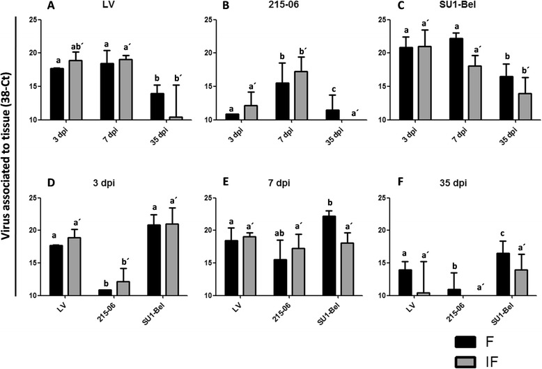
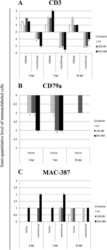
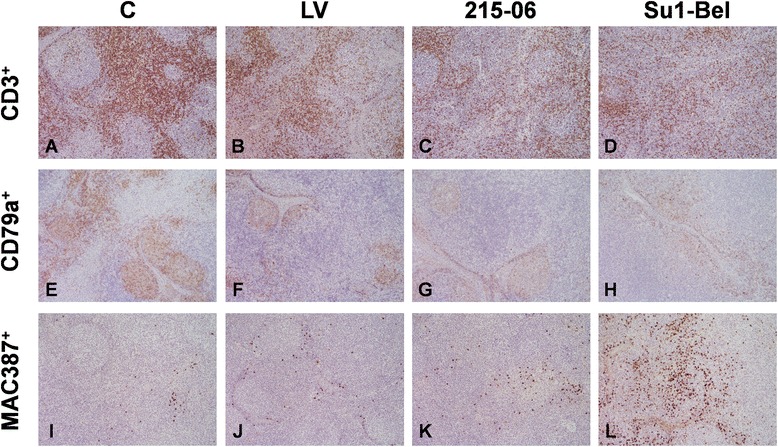
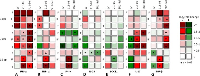
Porcine reproductive and respiratory syndrome virus (PRRSV) induces a weak immune response enabling it to persist in different organs of infected pigs. This has been attributed to the ability of PRRSV to influence the induction of cytokine responses. In this study, we investigated the cytokine transcriptional profiles in different compartments of the mediastinal lymph node of pigs infected with three genotype 1 PRRSV strains of differing pathogenicity: the low virulence prototype Lelystad virus (LV), and UK field strain 215-06 and the highly virulent subtype 3 SU1-Bel isolate from Belarus. We have used a combination of laser capture micro-dissection (LCM) followed by real time quantitative PCR (RT-qPCR) and immunohistochemical (IHC) detection of immune cell markers (CD3, CD79a and MAC387) and RT-qPCR quantification of PRRSV and cytokine transcripts. Compared to mock infected pigs, we found a significant downregulation of TNF-α and IFN-α in follicular and interfollicular areas of the mediastinal lymph node from 3 days post-infection (dpi) in animals infected with all three strains. This was accompanied by a transient B cell depletion and T cell and macrophage infiltration in the follicles together with T cell depletion in the interfollicular areas. A delayed upregulation of IFN-γ and IL-23p19 was observed mainly in the follicles. The PRRSV load was higher in all areas and time-points studied in the animals infected with the SU1-Bel strain. This paper describes the first application of LCM to study the cytokine transcript profiles and virus distribution in different compartments of the lymph node of pigs.


Figures

Figure 1
Figure 1
PRRSV viral load in mediastinal lymph node. PRRSV RNA was quantified by RT-qPCR and data represented by changes in the cycle threshold (Ct) in F and IF areas of Med-LN of LV (A), 215–06 (B) and SU1-Bel (C) infected animals. Differences between distinct PRRSV-1 infected groups are showed for 3 (D), 7 (E), and 35 (F) days post-infection. This figure showed the median of each group ± SD. In control animals the viral RNA were not detected (data not shown). Identical superscript letters indicate no significant difference (p > 0.05), whereas different superscript letters indicate statistically significant differences (p < 0.05) between the same compartment among different groups (a, b, and c for follicle; a´, b´, and c´ for interfollicular area).
Figure 2
Figure 2
Immunolabelled cell populations in mediastinal lymph node of PRRSV-1 infected pigs. A. CD3 staining for T cells detection; it was observed a general T cell depletion during this study for all PRRSV infected groups, otherwise the T cell immunolabeled cells increased in F at 7 dpi. B. CD79a staining for B cell staining in follicle, where was evidence a B cell depletion at 3 and 7 dpi for all PRRSV infected animals. C. Macrophage detection with MAC-387 staining, the amount of immunolabeled macrophages increased along this study in SU1-Bel infected pigs. The bars represent s-qic mean values; asterisks indicated the statistically significant differences (p < 0.05) against the control group. It is important to note that only a few animals in the control groups showed a higher number of immunolabeled cells in the follicle compared to the rest of animals within this group. For this reason there is a bar in some control groups.
Figure 3
Figure 3
Representative images of CD3, CD79a and MAC387 IHC staining in mediastinal lymph node. CD3 (A, B, C, D), CD79a (E, F, G, H) and MAC387 (I, J, K, L) in mediastinal lymph nodes of control pigs (A, E, I) and infected with LV (B, F, J), 215–06 (C, G, K) and SU1-Bel (D, H, J) strains, at 7 dpi. An increased in the number of CD3+ cells is observed in the lymphoid follicles from all infected groups together with a depletion of these cells in the interfollicular areas. A decrease in the number of CD79a cells in the follicles is also observed in all the infected groups. A substantial increase in the number of MAC387 is observed in the follicles and interfollicular areas from SU1-Bel infected animals (L) together with a mild increase in the LV (J) and 215–06 (K) groups. Original magnification: 20x.
Figure 4
Figure 4
Cytokine gene expression in mediastinal lymph node compartments. Log 2 Fold change in transcript level of IFN-α (A), TNF-α (B), IFN-γ (C), IL-23p19 (D), SOCS1 (E), IL-10 (F) and TGF-β (G) gene expression relative to the control gene β-Actin; which was calculated by 2-ΔΔCt method. Log2 fold change of gene expression in follicle (F), interfollicular area (IF) or total Med-LN lymph node (T) values are depicted. Green boxes indicate upregulation and red boxes indicate downregulation as the key showed. The statistically significant differences (p < 0.05) with control group are indicated by asterisks on the boxes.

Similar articles

Cited by

References

    1. Hopper SA, White ME, Twiddy N. An outbreak of blue-eared pig disease porcine reproductive and respiratory syndrome in four pig herds in Great Britain. Vet Rec. 1992;131:140–144. doi: 10.1136/vr.131.7.140. - DOI - PubMed
    1. Done SH, Paton DJ. Porcine reproductive and respiratory syndrome: clinical disease, pathology and immunosuppression. Vet Rec. 1995;136:32–35. doi: 10.1136/vr.136.2.32. - DOI - PubMed
    1. Rossow KD. Porcine reproductive and respiratory syndrome. Vet Pathol. 1998;35:1–20. doi: 10.1177/030098589803500101. - DOI - PubMed
    1. Zimmerman JJ, Benfield DA, Murtaugh MP, Osorio F, Stenvenson GW, Torremorell M (2006) Porcine reproductive and respiratory syndrome virus (porcine arterivirus). InStraw BE, Zimmerman JJ, D’Allaire S, Taylor DJ (eds) Ames, Iowa5. Holtkamp DJ, Kliebenstein JB, Neumann EJ, Zimmerman JJ, Rotto HF, Yoder TK, Wang C, Yeske PE, Mowrer CL, Haley CA (2013) Assessment of the economic impact of porcine reproductive and respiratory syndrome virus on United States pork producers. J Swine Health Prod 21:72–84
    1. Holtkamp DJ, Kliebenstein JB, Neumann EJ, Zimmerman JJ, Rotto HF, Yoder TK, Wang C, Yeske PE, Mowrer CL, Haley CA. Assessment of the economic impact of porcine reproductive and respiratory syndrome virus on United States pork producers. J Swine Health Prod. 2013;21:72–84.

Publication types

MeSH terms

LinkOut - more resources