

Histopathology and the inflammatory response of European perch, Perca fluviatilis muscle infected with Eustrongylides sp. (Nematoda)
- PMID: 25889096
- PMCID: PMC4404125
- DOI: 10.1186/s13071-015-0838-x
Histopathology and the inflammatory response of European perch, Perca fluviatilis muscle infected with Eustrongylides sp. (Nematoda)
Abstract
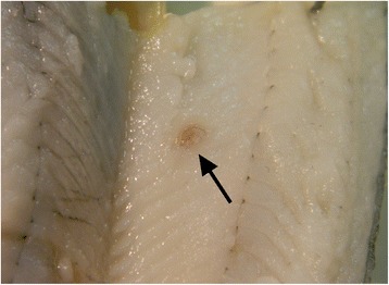
Background: The European perch, Perca fluviatilis L. is a common paratenic host of dioctophymatid nematodes belonging to the genus Eustrongylides. In this host, once infected oligochaetes, which serve as the first intermediate host, are ingested, Eustrongylides migrates through the intestine and is frequently encountered within the musculature, free within the body cavity, or encapsulated on the viscera. The current study details the first Italian record of Eustrongylides sp. with larvae reported in the muscle of P. fluviatilis.
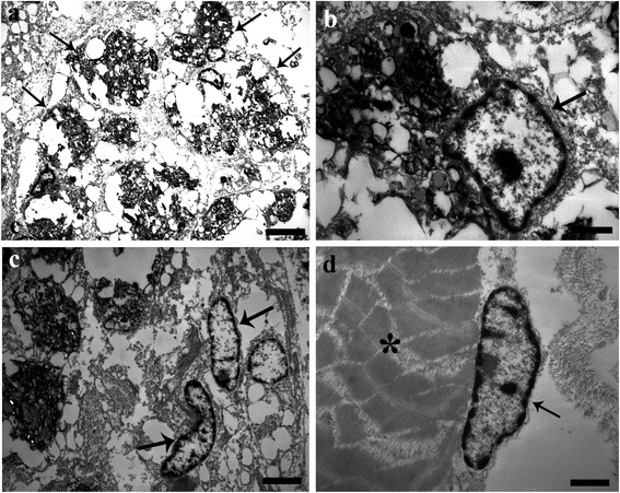
Methods: Uninfected and nematode-infected muscle tissues of perch were fixed and prepared for histological evaluation and electron microscopy. Some sections were subjected to an indirect immunohistochemical method using anti-PCNA, anti-piscidin 3 and anti-piscidin 4 antibodies.
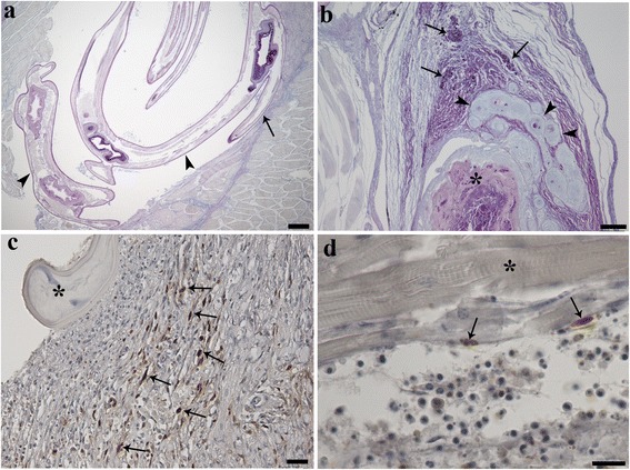
Results: A total of 510 P. fluviatilis (TL range 15-25 cm) from Lake Trasimeno, Perugia were post-mortemed; 31 individuals had encysted nematode larvae within their musculature (1-2 worms fish(-1)). Histologically, larvae were surrounded by a capsule with an evident acute inflammatory reaction. Muscle degeneration and necrosis extending throughout the sarcoplasm, sarcolemmal basal lamina, endomysial connective tissue cells and capillaries was frequently observed. Within the encapsulating reaction, macrophage aggregates (MAs) were seen. Immunohistochemical staining with the proliferating cell nuclear antigen (PCNA) revealed numerous PCNA-positive cells within the thickness of the capsule and in the immediate vicinity surrounding Eustrongylides sp. larvae (i.e. fibroblasts and satellite cells), suggesting a host response had been initiated to repair the nematode-damaged muscle. Mast cells (MCs) staining positively for piscidin 3, were demonstrated for the first time in response to a muscle-infecting nematode. The piscidin 3 positive MC's were seen principally in the periphery of the capsule surrounding the Eustrongylides sp. larva.
Conclusions: A host tissue response to Eustrongylides sp. larvae infecting the musculature of P. fluviatilis was observed. Numerous fibroblasts, MAs and MCs were seen throughout the thick fibroconnectival layer of the capsule enclosing larvae. PCNA positive cells within the capsule suggest that host repair of nematode damaged muscle does occur, while the presence of the antimicrobial peptide piscidin 3 is shown for the first time. This is first report of Eustrongylides sp. in an Italian population of P. fluviatilis.
Figures
References
-
- Moravec F. Parasitic Nematodes of Freshwater Fishes of Europe. Revised Second Edition. Hardback, Academia: Praha; 2013.
Publication types
MeSH terms
LinkOut - more resources
Full Text Sources
Other Literature Sources
Medical
Miscellaneous
