

Animal model of acute gout reproduces the inflammatory and ultrasonographic joint changes of human gout
- PMID: 25889158
- PMCID: PMC4363186
- DOI: 10.1186/s13075-015-0550-4
Animal model of acute gout reproduces the inflammatory and ultrasonographic joint changes of human gout
Abstract
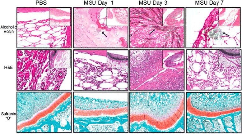
Introduction: Gout is an inflammatory condition induced by the deposition of monosodium urate (MSU) crystals in the joints and soft tissues that can produce acute or chronic arthritis. Several animal models of crystal-induced inflammation have been proposed that involve direct injection of MSU-crystals into different anatomical structures; however, only a few of these models reflect a true diarthrodial joint microenvironment in which an acute gouty attack takes place. The aim of this study was to assess the inflammatory and structural joint changes in a rabbit model of acute gout attack by ultrasound (US), synovial fluid (SF) and histopathological analyses.
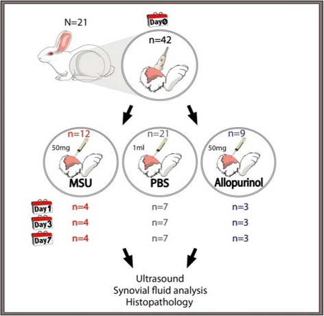
Methods: Under US guidance, 42 rabbit knees were randomly injected with a suspension of 50 mg/ml of either MSU or allopurinol synthetic crystals. The control group received intra-articular vehicle of phosphate-buffered saline (PBS). US evaluation, SF and histopathological analyses were performed at days 1, 3, and 7.
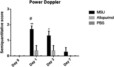
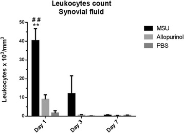
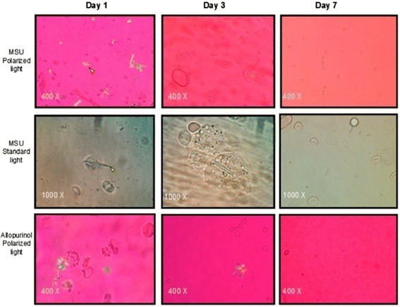
Results: A total of 21 rabbit knees were assigned to the control group, 12 to the MSU-crystals group, and 9 to the allopurinol crystals group. By US, the MSU crystals group displayed the double contour sign and bright stippled aggregates in 67% and 75% of joints, respectively. Neither control knees nor allopurinol crystals group displayed these US signs. Power Doppler (PD) signal was moderate to intense in the MSU-crystals group and greater than both the allopurinol crystal and control groups at day 1 (P<0.001) and 3 (P<0.05), with its practical disappearance by day 7. SF leukocyte count was 40,312±6,369 cells/mm3 in the MSU-crystals group, higher than in controls (P=0.004) and allopurinol crystal group (P=0.006). At day 7, SF leukocyte count decreased in both MSU and allopurinol crystal groups reaching the non-inflammatory range. Histologically, at day 3 intense synovial polymorphonuclear cells infiltration and MSU aggregates were identified.
Conclusion: The rabbit model of MSU crystal-induced acute arthritis efficiently reproduces the inflammatory, US, SF and histopathological changes of the human acute gouty attack.
Figures

Similar articles
-
Intra-articular corticosteroid preparations: different characteristics and their effect during inflammation induced by monosodium urate crystals in the rat subcutaneous air pouch.Rheumatology (Oxford). 2003 Sep;42(9):1093-100. doi: 10.1093/rheumatology/keg305. Epub 2003 May 30. Rheumatology (Oxford). 2003. PMID: 12777646
-
Validation of Musculoskeletal Ultrasound in the Assessment of Experimental Gout Synovitis.Ultrasound Med Biol. 2018 Jul;44(7):1516-1524. doi: 10.1016/j.ultrasmedbio.2018.03.018. Epub 2018 Apr 24. Ultrasound Med Biol. 2018. PMID: 29703511
-
Diagnostic value of ultrasound versus dual-energy computed tomography in patients with different stages of acute gouty arthritis.Clin Rheumatol. 2020 May;39(5):1649-1653. doi: 10.1007/s10067-020-05014-6. Epub 2020 Mar 10. Clin Rheumatol. 2020. PMID: 32157468
-
Clinical features of gout.Reumatismo. 2012 Jan 19;63(4):238-45. doi: 10.4081/reumatismo.2011.238. Reumatismo. 2012. PMID: 22303530 Review.
-
P2X7R: a potential key regulator of acute gouty arthritis.Semin Arthritis Rheum. 2013 Dec;43(3):376-80. doi: 10.1016/j.semarthrit.2013.04.007. Epub 2013 Jun 17. Semin Arthritis Rheum. 2013. PMID: 23786870 Review.
Cited by
-
A fly GWAS for purine metabolites identifies human FAM214 homolog medusa, which acts in a conserved manner to enhance hyperuricemia-driven pathologies by modulating purine metabolism and the inflammatory response.Geroscience. 2022 Aug;44(4):2195-2211. doi: 10.1007/s11357-022-00557-9. Epub 2022 Apr 6. Geroscience. 2022. PMID: 35381951 Free PMC article.
-
Performance of Ultrasound in the Clinical Evaluation of Gout and Hyperuricemia.J Immunol Res. 2021 Apr 5;2021:5550626. doi: 10.1155/2021/5550626. eCollection 2021. J Immunol Res. 2021. PMID: 33884273 Free PMC article.
-
MSC therapy ameliorates experimental gouty arthritis hinting an early COX-2 induction.Front Immunol. 2023 Jul 18;14:1193179. doi: 10.3389/fimmu.2023.1193179. eCollection 2023. Front Immunol. 2023. PMID: 37533852 Free PMC article.
-
Anti-Inflammatory and Hypouricemic Effect of Bioactive Compounds: Molecular Evidence and Potential Application in the Management of Gout.Curr Issues Mol Biol. 2022 Oct 25;44(11):5173-5190. doi: 10.3390/cimb44110352. Curr Issues Mol Biol. 2022. PMID: 36354664 Free PMC article. Review.
-
Case Report: Articular Gout in Four Dogs and One Cat.Front Vet Sci. 2022 Apr 26;9:752774. doi: 10.3389/fvets.2022.752774. eCollection 2022. Front Vet Sci. 2022. PMID: 35558881 Free PMC article.
References
Publication types
MeSH terms
Substances
Grants and funding
LinkOut - more resources
Full Text Sources
Other Literature Sources
Medical
