

Lung segmentation on standard and mobile chest radiographs using oriented Gaussian derivatives filter
- PMID: 25889188
- PMCID: PMC4355502
- DOI: 10.1186/s12938-015-0014-8
Lung segmentation on standard and mobile chest radiographs using oriented Gaussian derivatives filter
Abstract
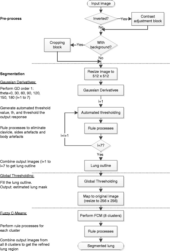
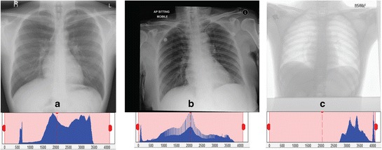
Background: Unsupervised lung segmentation method is one of the mandatory processes in order to develop a Content Based Medical Image Retrieval System (CBMIRS) of CXR. The purpose of the study is to present a robust solution for lung segmentation of standard and mobile chest radiographs using fully automated unsupervised method.
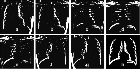
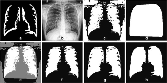
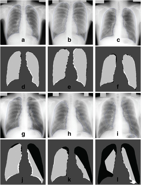
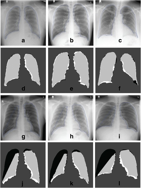
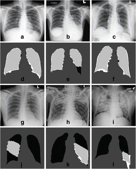
Methods: The novel method is based on oriented Gaussian derivatives filter with seven orientations, combined with Fuzzy C-Means (FCM) clustering and thresholding to refine the lung region. In addition, a new algorithm to automatically generate a threshold value for each Gaussian response is also proposed. The algorithms are applied to both PA and AP chest radiographs from both public JSRT dataset and our private datasets from collaborative hospital. Two pre-processing blocks are introduced to standardize the images from different machines. Comparisons with the previous works found in the literature on JSRT dataset shows that our method gives a reasonably good result. We also compare our algorithm with other unsupervised methods to provide fairly comparative measures on the performances for all datasets.
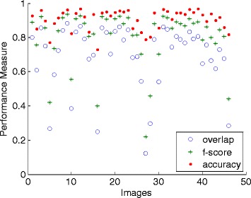
Results: Performance measures (accuracy, F-score, precision, sensitivity and specificity) for the segmentation of lung in public JSRT dataset are above 0.90 except for the overlap measure is 0.87. The standard deviations for all measures are very low, from 0.01 to 0.06. The overlap measure for the private image database is 0.81 (images from standard machine) and 0.69 (images from two mobile machines). The algorithm is fully automated and fast, with the average execution time of 12.5 s for 512 by 512 pixels resolution.
Conclusions: Our proposed method is fully automated, unsupervised, with no training or learning stage is necessary to segment the lungs taken using both a standard machine and two different mobile machines. The proposed pre-processing blocks are significantly useful to standardize the radiographs from mobile machines. The algorithm gives good performance measures, robust, and fast for the application of the CBMIRS.
Figures


Similar articles
-
A review on lung boundary detection in chest X-rays.Int J Comput Assist Radiol Surg. 2019 Apr;14(4):563-576. doi: 10.1007/s11548-019-01917-1. Epub 2019 Feb 7. Int J Comput Assist Radiol Surg. 2019. PMID: 30730032 Free PMC article. Review.
-
A novel fuzzy C-means algorithm for unsupervised heterogeneous tumor quantification in PET.Med Phys. 2010 Mar;37(3):1309-24. doi: 10.1118/1.3301610. Med Phys. 2010. PMID: 20384268
-
Unsupervised segmentation of lung fields in chest radiographs using multiresolution fractal feature vector and deformable models.Med Biol Eng Comput. 2016 Sep;54(9):1409-22. doi: 10.1007/s11517-015-1412-6. Epub 2015 Nov 3. Med Biol Eng Comput. 2016. PMID: 26530048
-
Brain tumor segmentation approach based on the extreme learning machine and significantly fast and robust fuzzy C-means clustering algorithms running on Raspberry Pi hardware.Med Hypotheses. 2020 Mar;136:109507. doi: 10.1016/j.mehy.2019.109507. Epub 2019 Nov 18. Med Hypotheses. 2020. PMID: 31812927
-
Recent computational methods for white blood cell nuclei segmentation: A comparative study.Comput Methods Programs Biomed. 2019 May;173:1-14. doi: 10.1016/j.cmpb.2019.03.001. Epub 2019 Mar 6. Comput Methods Programs Biomed. 2019. PMID: 31046984 Review.
Cited by
-
CardioNet: Automatic Semantic Segmentation to Calculate the Cardiothoracic Ratio for Cardiomegaly and Other Chest Diseases.J Pers Med. 2022 Jun 17;12(6):988. doi: 10.3390/jpm12060988. J Pers Med. 2022. PMID: 35743771 Free PMC article.
-
A review on lung boundary detection in chest X-rays.Int J Comput Assist Radiol Surg. 2019 Apr;14(4):563-576. doi: 10.1007/s11548-019-01917-1. Epub 2019 Feb 7. Int J Comput Assist Radiol Surg. 2019. PMID: 30730032 Free PMC article. Review.
-
Computer-aided detection in chest radiography based on artificial intelligence: a survey.Biomed Eng Online. 2018 Aug 22;17(1):113. doi: 10.1186/s12938-018-0544-y. Biomed Eng Online. 2018. PMID: 30134902 Free PMC article. Review.
-
Segmentation of lung fields from chest radiographs-a radiomic feature-based approach.Biomed Eng Lett. 2018 Oct 17;9(1):109-117. doi: 10.1007/s13534-018-0086-z. eCollection 2019 Feb. Biomed Eng Lett. 2018. PMID: 30956884 Free PMC article.
-
A deep learning based dual encoder-decoder framework for anatomical structure segmentation in chest X-ray images.Sci Rep. 2023 Jan 16;13(1):791. doi: 10.1038/s41598-023-27815-w. Sci Rep. 2023. PMID: 36646735 Free PMC article.
References
-
- Goldstone K, Yates SJ. Radiation issues governing radiation protection and patient doses in diagnostic imaging. In: Adam A, Dixon AK, editors. Grainger & Allison’s diagnostic radiology. New York: Churchill Livingstone; 2008.
Publication types
MeSH terms
Substances
LinkOut - more resources
Full Text Sources
Other Literature Sources
