Follicle dynamics and global organization in the intact mouse ovary
- PMID: 25889274
- PMCID: PMC4469539
- DOI: 10.1016/j.ydbio.2015.04.006
Follicle dynamics and global organization in the intact mouse ovary
Abstract
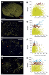
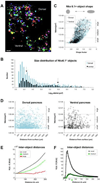
Quantitative analysis of tissues and organs can reveal large-scale patterning as well as the impact of perturbations and aging on biological architecture. Here we develop tools for imaging of single cells in intact organs and computational approaches to assess spatial relationships in 3D. In the mouse ovary, we use nuclear volume of the oocyte to read out quiescence or growth of oocyte-somatic cell units known as follicles. This in-ovary quantification of non-growing follicle dynamics from neonate to adult fits a mathematical function, which corroborates the model of fixed oocyte reserve. Mapping approaches show that radial organization of folliculogenesis established in the newborn ovary is preserved through adulthood. By contrast, inter-follicle clustering increases during aging with different dynamics depending on size. These broadly applicable tools can reveal high dimensional phenotypes and age-related architectural changes in other organs. In the adult mouse pancreas, we find stochastic radial organization of the islets of Langerhans but evidence for localized interactions among the smallest islets.
Keywords: Confocal imaging; Islet of Langerhans; Ovary; Pancreas; Primordial follicle; Whole tissue labeling.
Copyright © 2015 Elsevier Inc. All rights reserved.
Figures


Similar articles
-
KIT/KIT ligand in mammalian oogenesis and folliculogenesis: roles in rabbit and murine ovarian follicle activation and oocyte growth.Biol Reprod. 2006 Sep;75(3):421-33. doi: 10.1095/biolreprod.106.051516. Epub 2006 Jun 21. Biol Reprod. 2006. PMID: 16790689
-
The ontogeny of primordial follicles in the mouse ovary.Prog Clin Biol Res. 1989;296:55-63. Prog Clin Biol Res. 1989. PMID: 2740401 No abstract available.
-
Three-dimensional localization and dynamics of centromeres in mouse oocytes during folliculogenesis.J Mol Histol. 2004 Aug;35(6):631-8. doi: 10.1007/s10735-004-2190-x. J Mol Histol. 2004. PMID: 15614617
-
Driving folliculogenesis by the oocyte-somatic cell dialog: Lessons from genetic models.Theriogenology. 2016 Jul 1;86(1):41-53. doi: 10.1016/j.theriogenology.2016.04.017. Epub 2016 Apr 21. Theriogenology. 2016. PMID: 27155734 Review.
-
The ovarian reserve of primordial follicles and the dynamic reserve of antral growing follicles: what is the link?Biol Reprod. 2014 Apr 25;90(4):85. doi: 10.1095/biolreprod.113.117077. Print 2014 Apr. Biol Reprod. 2014. PMID: 24599291 Review.
Cited by
-
Hyperandrogenism triggers mtDNA release to participate in ovarian inflammation via mPTP/cGAS/STING in PCOS.iScience. 2025 Apr 8;28(5):112391. doi: 10.1016/j.isci.2025.112391. eCollection 2025 May 16. iScience. 2025. PMID: 40322081 Free PMC article.
-
Ovary Development: Insights From a Three-Dimensional Imaging Revolution.Front Cell Dev Biol. 2021 Jul 26;9:698315. doi: 10.3389/fcell.2021.698315. eCollection 2021. Front Cell Dev Biol. 2021. PMID: 34381780 Free PMC article. Review.
-
A Roadmap for Three-Dimensional Analysis of the Intact Mouse Ovary.Methods Mol Biol. 2023;2677:203-219. doi: 10.1007/978-1-0716-3259-8_12. Methods Mol Biol. 2023. PMID: 37464244 Free PMC article.
-
Comparison of methods for quantifying primordial follicles in the mouse ovary.J Ovarian Res. 2020 Oct 14;13(1):121. doi: 10.1186/s13048-020-00724-6. J Ovarian Res. 2020. PMID: 33054849 Free PMC article.
-
Tissue clearing and imaging approaches for in toto analysis of the reproductive system†.Biol Reprod. 2024 Jun 12;110(6):1041-1054. doi: 10.1093/biolre/ioad182. Biol Reprod. 2024. PMID: 38159104 Free PMC article. Review.
References
-
- Bristol-Gould SK, Kreeger PK, Selkirk CG, Kilen SM, Cook RW, Kipp JL, Shea LD, Mayo KE, Woodruff TK. Postnatal regulation of germ cells by activin: The establishment of the initial follicle pool. Dev. Biol. 2006a;298(1):132–148. - PubMed
-
- Bristol-Gould SK, Kreeger PK, Selkirk CG, Kilen SM, Mayo KE, Shea LD, Woodruff TK. Fate of the initial follicle pool: Empirical and mathematical evidence supporting its sufficiency for adult fertility. Dev. Biol. 2006b;298(1):149–154. - PubMed
-
- Byskov AG, Guoliang X, Andersen CY. The cortex-medulla oocyte growth pattern is organized during fetal life: an in-vitro study of the mouse ovary. Mol. Hum. Reprod. 1997;3(9):795–800. - PubMed
Publication types
MeSH terms
Grants and funding
LinkOut - more resources
Full Text Sources
Other Literature Sources

