Review
. 2015 Apr 8;15(1):5.
doi: 10.1186/s40644-015-0040-6.

Neuroblastoma and nephroblastoma: a radiological review

Affiliations
Review

Neuroblastoma and nephroblastoma: a radiological review

Maureen Dumba et al. Cancer Imaging. .

Abstract

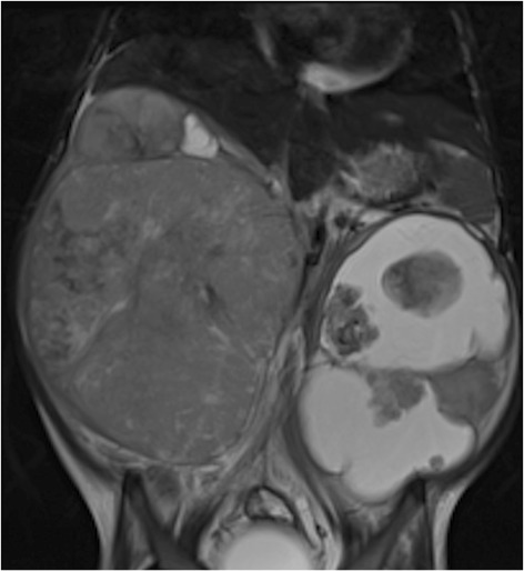
Neuroblastoma (NBL) is the most common extra-cranial tumour in childhood. It can present as an abdominal mass, but is usually metastatic at diagnosis so the symptomatology can be varied. Nephroblastoma, also more commonly known as a Wilms tumour, is the commonest renal tumour in childhood and more typically presents as abdominal pathology with few constitutional symptoms, although rarely haematuria can be a presenting feature. The pathophysiology and clinical aspects of both tumours including associated risk factors and pathologies are discussed. Oncogenetics and chromosomal abnormalities are increasingly recognised as important prognostic indicators and their impact on initial management is considered. Imaging plays a pivotal role in terms of diagnosis and recent imaging advances mean that radiology has an increasingly crucial role in the management pathway. The use of image defined risk factors in neuroblastoma has begun to dramatically change how this tumour is characterised pre-operatively. The National Wilms Tumour Study Group have comprehensively staged Wilms tumours and this is reviewed as it impacts significantly on management. The use of contrast-enhanced MRI and diffusion-weighted sequences have further served to augment the information available to the clinical team during initial assessment of both neuroblastomas and Wilms tumours. The differences in management strategies are outlined. This paper therefore aims to provide a comprehensive update on these two common paediatric tumours with a particular emphasis on the current crucial role played by imaging.


Figures

Figure 1
Figure 1
Chest x-ray of 3 year old girl showing thoracic NBL. Note erosions of the posterior 3 rd and 4 th ribs indicating a posterior mediastinal mass.
Figure 2
Figure 2
Coronal T2 MR of a 3 year old boy with extensive abdominal NBL that crosses the midline and is here seen to encase the aorta (blue arrow).
Figure 3
Figure 3
Axial T2 MR of 2 year old girl showing NBL with rib invasion (blue arrow), anterior aortic displacement and encasement (red arrow) and bilateral pleural effusions.
Figure 4
Figure 4
Axial T2 MR of 3 year old boy showing intraspinal extent of NBL with tumour seen in both neural foramina on this single image (blue arrows).
Figure 5
Figure 5
Coronal T2 MR of a 2 year old boy showing left-sided NBL mass with bone marrow involvement (blue arrow).
Figure 6
Figure 6
Axial CT of a 2 year old girl showing a left-sided abdominal NBL with evidence of calcification (blue arrows).
Figure 7
Figure 7
MIBG scintigraphy showing avid uptake at the site of the abdominal NBL with widespread bony metastatic spread.
Figure 8
Figure 8
Axial T2 MR of a 4 year old boy with right-sided Wilms tumour and left-sided nephroblastomatosis (blue arrow).
Figure 9
Figure 9
Ultrasound abdomen of a 4 year old boy with a left-sided Wilms tumour, presenting here unusually as a solid uniform mass.
Figure 10
Figure 10
Coronal T2 fat-saturated MR of 3 year old boy showing a right sided Wilms tumour with the ‘claw sign’ of normal renal tissue (blue arrow) surrounding the tumour.
Figure 11
Figure 11
Coronal T2 MR of a 4 year old girl with bilateral Wilms tumours, more cystic on the left.
Figure 12
Figure 12
Axial CT of a 3 year old boy with right-sided Wilms tumour, again demonstrating the ‘claw sign’ (blue arrow).

References

    1. Maris JM, Hogarty MD, Bagatell R, Cohn SL. Neuroblastoma. Lancet. 2007;369:2106–20. doi: 10.1016/S0140-6736(07)60983-0. - DOI - PubMed
    1. Xu Y, Wang J, Peng Y, Zeng J. CT characteristics of primary retroperitoneal neoplasms in children. Eur J Rad. 2010;75:321–328. doi: 10.1016/j.ejrad.2010.05.041. - DOI - PubMed
    1. Hiorns M, Owens CM. Radiology of neuroblastoma in paediatrics. Eur Radiol. 2001;11:2071–81. doi: 10.1007/s003300100931. - DOI - PubMed
    1. Ora I, Eggert A. Progress in treatment and risk stratification of neuroblastoma: Impact on future clinical and basic research. Semi Cancer Biol. 2001;21:217–28. doi: 10.1016/j.semcancer.2011.07.002. - DOI - PubMed
    1. Survival rates for neuroblastoma. [http://www.cancer.org/cancer/neuroblastoma/detailedguide/neuroblastoma-s...]