Clinical Trial
. 2015 Jan 27:13:17.
doi: 10.1186/s12967-014-0371-2.

Human neural stem cell transplantation in ALS: initial results from a phase I trial

Affiliations
Clinical Trial

Human neural stem cell transplantation in ALS: initial results from a phase I trial

Letizia Mazzini et al. J Transl Med. .

Abstract

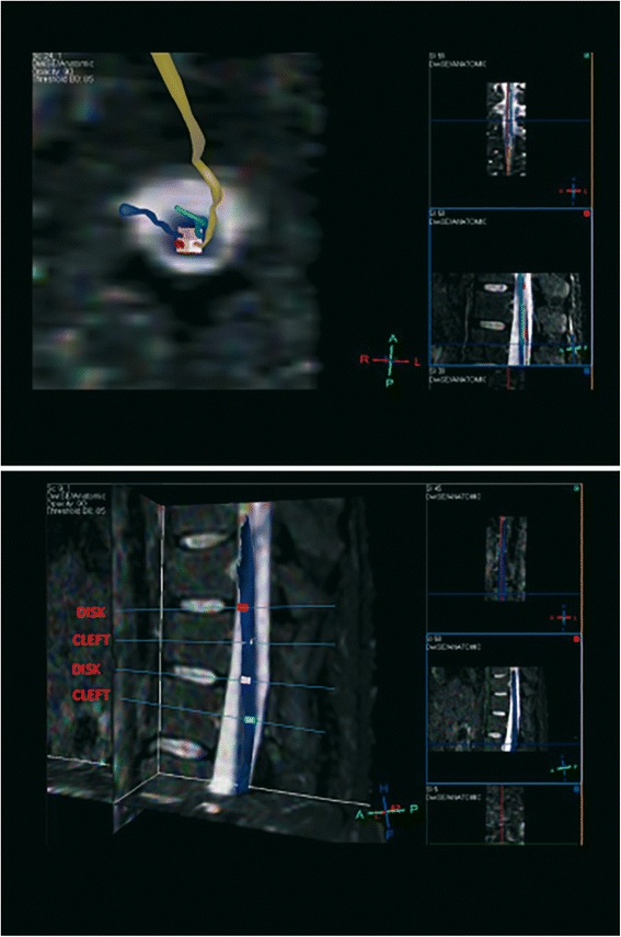
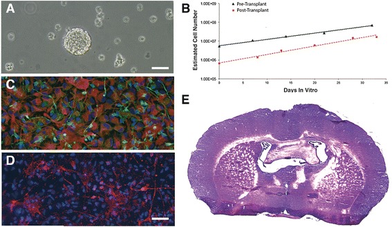
Background: We report the initial results from a phase I clinical trial for ALS. We transplanted GMP-grade, fetal human neural stem cells from natural in utero death (hNSCs) into the anterior horns of the spinal cord to test for the safety of both cells and neurosurgical procedures in these patients. The trial was approved by the Istituto Superiore di Sanità and the competent Ethics Committees and was monitored by an external Safety Board.

Methods: Six non-ambulatory patients were treated. Three of them received 3 unilateral hNSCs microinjections into the lumbar cord tract, while the remaining ones received bilateral (n = 3 + 3) microinjections. None manifested severe adverse events related to the treatment, even though nearly 5 times more cells were injected in the patients receiving bilateral implants and a much milder immune-suppression regimen was used as compared to previous trials.

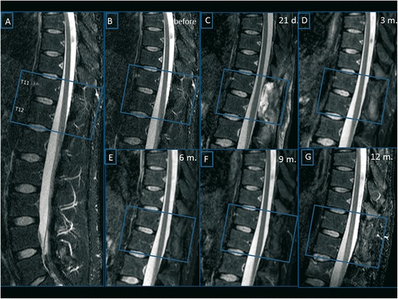
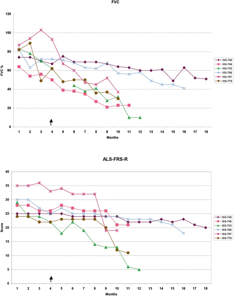
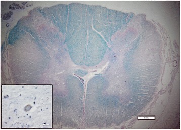
Results: No increase of disease progression due to the treatment was observed for up to18 months after surgery. Rather, two patients showed a transitory improvement of the subscore ambulation on the ALS-FRS-R scale (from 1 to 2). A third patient showed improvement of the MRC score for tibialis anterior, which persisted for as long as 7 months. The latter and two additional patients refused PEG and invasive ventilation and died 8 months after surgery due to the progression of respiratory failure. The autopsies confirmed that this was related to the evolution of the disease.

Conclusions: We describe a safe cell therapy approach that will allow for the treatment of larger pools of patients for later-phase ALS clinical trials, while warranting good reproducibility. These can now be carried out under more standardized conditions, based on a more homogenous repertoire of clinical grade hNSCs. The use of brain tissue from natural miscarriages eliminates the ethical concerns that may arise from the use of fetal material.

Trial registration: EudraCT:2009-014484-39 .


Figures

Figure 1
Figure 1
Spinal cord MRI. Upper Panel: Diffusion Tensor Imaging (DTI) overlaid with the STIR pulse sequence, in a MPR algorithm. For each of single point (colored box) ROIs, a correspondent fiber is evoked and reconstructed to ensure that the level examined and the ROI adopted is inside the spinal cord. Lower Panel: DTI post-processing by mean of a MPR algorithm, with overlaying of the STIR pulse sequence, to select the exact levels to be studied at this time of examination and in further MR follow-up scans. Disk and somatic vascular cleft are adopted to select the proper planes.
Figure 2
Figure 2
Cell quality control. (A) Growth kinetics of a set of hNSCs lines showing the increasing, estimated overall cell number at each passage. (B) Clonal efficiency assay showing the percentage of cells that retain the ability to form clonal neurospheres over the total cell number plated is reported (Replicates n = 3), bars describe standard error. (C) hNSCs differentiate into astrocytes (left, green, GFAP), neurons (left, red, βIII-tubulin) and oligodendrocytes (right, GalC, red); nuclei are counterstained in blue (DAPI). (Bar = 50 μm). (D) All of the hNSCs lines tested undergo extinction in vitro upon growth factor removal, as shown by the negative growth kinetic in which the total cell number approaches zero in a few passages.
Figure 3
Figure 3
The cell lines used in this study were confirmed to retain a normal karyotype all throughout passaging. The figure shows the example of a karyogram performed on the hNSC line from a female donor (46, XX) after seventeen passages. Chromosome G-banding was routinely performed on both the Intermediate Product and the Finished Product. In addition we also tested for karyotype stability the cells that were left in the needle, post-transplantation.
Figure 4
Figure 4
hNSCs transplant into Nude Mice CNS. The lateral striatum of nude mice was the target area (A, arrow) for the transplantation of normal hNSCs (B) or glioblastoma cancer stem cells (GBM; positive graft controls; C). Mice were sacrificed six (B) and two months (C) after transplantation, respectively. The hematoxilyn/eosyn stain showed that structural organization of the transplanted regions was well preserved in mice tranplanted with hNSCs (B), whereas hypercellularity and anomalous growth and necrosis ensued in regions receiving GBM cells (C). Confocal microscopy of anti-human nuclei staining (huN, green, D) showed that hNSCs engrafted efficiently, with only a few human cells retaining residual proliferation activity as shown by co-labeling with the proliferation marker ki67 (red). hNSCs labeled with huN (E, green) differentiate into βTubIII+ neurons (E, red) and GFAP+ astrocytes (F, red). Nuclei are shown by DAPI staining. Scale bars: D-E = 15 μm; all insets: 10 μm, bar in inset D.
Figure 5
Figure 5
MRI Follow-up. T2 weighted sequences acquired on sagittal plane before surgery (images A-B) and respectively 21 days (image C), 3 (image D), 6 (image E), 9 (image F) and 12 months (image G) after transplantation. Post-surgical MR scans revealed an expected extradural fluid collection at the site of surgery, which resolved spontaneously. No structural changes were detected after hNSCs transplantation relative to the baseline.
Figure 6
Figure 6
Post Transplant hNSCs Test. (A) an example of human neural stem cells that were leftover from the transplant and put back in culture were they re-established typical neurosphere, expanding lines, with a growth profile that mirrored that of the very same cultures prior to the transplant, as shown in B. These cells differentiated into neurons (βIII-Tubulin, green, C) and astrocytes (GFAP, Red, C) and oligodendrocytes (GalC, Red, D). E: an example of whole brain reconstruction from one out of 10 nude mice that were transplanted into the right lateral striatum with the cells recovered from the transplant and recultured, showing no hyperplastic areas or tumor formation. Bars: A, 100 μm C,D 50 μm, Bar in D.
Figure 7
Figure 7
Clinical follow-up. Changes of the Forced Vital Capacity (upper panel) and of the ALS-FRS score (lower panel) in the 3-month period of natural history observation and after transplantation. The arrow indicates the time of NSCs transplantation.
Figure 8
Figure 8
Representative cross section of the spinal cord in the region of transplantation stained with luxol fast blue and periodic acid Schiff. There is no apparent disruption of tissue due to injection. Note the degeneration of the cortico-spinal tracts (“lateral sclerosis”). The inset demonstrates a phosphorylated TDP43 inclusion in a remaining motor neuron. Scale bars are 1 mm for the low power and 20 microns for the inset.

References

    1. Pluchino S, Cossetti C. How stem cells speak with host immune cells in inflammatory brain diseases. Glia. 2013;61(9):1379–40. doi: 10.1002/glia.22500. - DOI - PMC - PubMed
    1. Giusto E, Donegà M, Cossetti C, Pluchino S. Neuro-immune interactions of neural stem cell transplants: From animal disease models to human trials. Exp Neurol. 2013;16:S0014–4886(13). - PMC - PubMed
    1. Mazzini L, Vercelli A, Ferrero I, Mareschi K, Boido M, Servo S, et al. Stem cells in amyotrophic lateral sclerosis: state of the art. Expert Opin Biol Ther. 2009;9(10):1245–1258. doi: 10.1517/14712590903186956. - DOI - PubMed
    1. Mazzini L, Ferrero I, Luparello V, Rustichelli D, Gunetti M, Mareschi K, et al. Mesenchymal stem cell transplantation in amyotrophic lateral sclerosis: A Phase I clinical trial. Exp Neurol. 2010;223(1):229–237. doi: 10.1016/j.expneurol.2009.08.007. - DOI - PubMed
    1. Mazzini L, Mareschi K, Ferrero I, Miglioretti M, Stecco A, Servo S, et al. Mesenchymal stromal cell transplantation in amyotrophic lateral sclerosis: a long-term safety study. Cytotherapy. 2012;14(1):56–60. doi: 10.3109/14653249.2011.613929. - DOI - PubMed

Publication types

MeSH terms

Substances