Review
. 2015 Feb 22:13:13.
doi: 10.1186/s12964-015-0092-z.

Interleukin-1 loop model for pathogenesis of Langerhans cell histiocytosis

Affiliations
Review

Interleukin-1 loop model for pathogenesis of Langerhans cell histiocytosis

Ichiro Murakami et al. Cell Commun Signal. .

Abstract

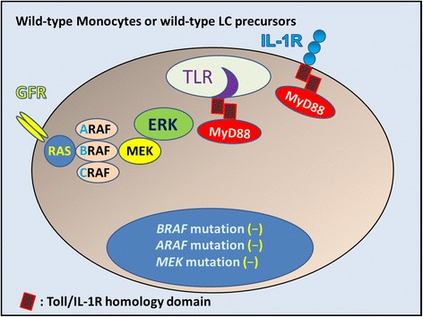
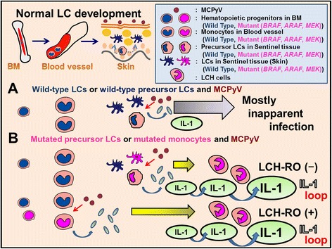
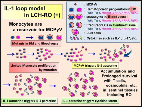
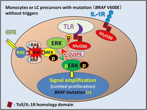
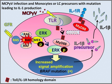
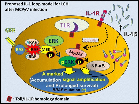
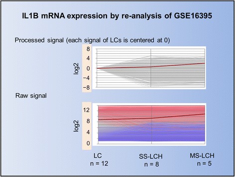
We propose Langerhans cell histiocytosis (LCH) is an inflammatory process that is prolonged by mutations. We hypothesize that Merkel cell polyomavirus (MCPyV) infection triggers an interleukin-1 (IL-1) activation loop that underlies the pathogenesis of LCH. Langerhans cells (LCs) are antigen presenting cells in the skin. When LCs encounter exogenous antigens, they migrate from the epidermis into draining lymphoid tissues to initiate T-cell activity. It has been proposed that LC migration-related factors, including E-cadherin, matrix metalloproteinase, and Notch ligand induce LCH activity. We found that the tyrosine phosphatase SHP-1, which binds IL-1 receptor-associated kinase 1, is expressed at a significantly higher level in LCH affecting multiple organ systems (MS-LCH) than in LCH affecting a single organ system (SS-LCH). IL-1 stimulates T helper 17 cells and their signature cytokine IL-17 had been a matter of controversy. We detected higher levels of IL-17A receptor expression in MS-LCH than in SS-LCH and proposed an IL-17 endocrine model that could settle the controversy. IL-1 is the first cytokine secreted in response to sensitizers and promotes LC migration from sentinel tissues. Myeloid differentiation primary response 88 (MyD88), downstream of the IL-1 receptor, has functions in both RAS signaling and inflammation, leading to human cell transformation. In 2010, an activating mutation in the B-rapidly accelerated fibrosarcoma gene (BRAF) V600E was found in LCH. This BRAF mutation induces phosphorylation of the extracellular signal-regulated kinase (ERK) that may play an important role with MyD88 in LCH pathogenesis. However, phosphorylated ERK (pERK) is rapidly dephosphorylated by dual specificity phosphatase 6 (DUSP6), and limited proliferation is predicted in BRAF mutant cells. MyD88 binds pERK via its D-domain, thereby preventing pERK-DUSP6 interaction and maintaining ERK in an active, phosphorylated state. We detected MCPyV-DNA in the peripheral blood cells of two out of three patients with LCH in high-risk organs but not in those of patients with LCH in non-high-risk organs (0/12; P = .029). MCPyV infection can trigger precursor LCH cells with BRAF mutation to produce IL-1; the IL-1 loop is amplified in all LCH subclasses. Our model indicates both BRAF mutation and IL-1 loop regulation as potential therapeutic targets.


Figures

Figure 1
Figure 1
Langerhans cells and Merkel cells in the epidermis. (A) Skin of one-year-old female baby with nevus cell nevus. (Hematoxylin and Eosin (H&E) stain). (B) Immunohistochemistry shows S100+ immature Langerhans cell (LC; red), nevus cell (NC; red), and cytokeratin 20 (CK20) + Merkel cells (MC; brown). Same area of panel A. (C) S100+ immature LC and NC (red) and CK20+ MC (brown) in a hair follicle. High power view of the left central part of panel B. (D) Immunohistochemistry shows CD1a + LC (blue) and CD1a- NC. Brown pigments correspond to melanin. Same area of panel A. (E) Immature LCs in the epidermis. High power view of panel D. (F) Immature LCs in a hair follicle are positive for CD1a, while the NCs are negative for CD1a. High power view of panel D.
Figure 2
Figure 2
Wild-type monocytes, wild-type LC precursors, or wild-type immature Langerhans cells without mutations. In these cells, mitogens such as growth factors bind to and activate cell-surface receptors (GFR: growth factor receptor) that induce signaling through a complex consisting of adaptor proteins and exchange factors to activate RAS (Blue circle) on the inner surface of the cell membrane. Once activated, RAS binds to and activates the RAF family of proteins, including BRAF, which subsequently phosphorylates and activates MEK. Activated MEK subsequently phosphorylates and activates ERK. Activated ERK phosphorylates numerous substrates within the cytoplasm and nucleus, promoting cell division and enhancing survival, movement, and differentiation. In the case of Merkel cell polyomavirus (MCPyV) infection, some LCs may present MCPyV antigen, inducing adaptive immunity through Toll-like receptors (TLRs). IL-1 is the first cytokine secreted in response to sensitizers. IL-1 binds to IL-1 receptor (IL-1R) and promotes LC migration from sentinel tissue such as the skin. MCPyV interferes with LC function and maturation to evade immune surveillance, which might allow infection by inhibiting NF-κB essential modulator (NEMO) and down-regulation of TLR9.
Figure 3
Figure 3
Proposed relationship between MCPyV and wild-type Langerhans cell (LC) precursors and wild-type LCs or mutated monocytes and mutated LC precursors. (A) MCPyV usually causes inapparent infection with immunoglobulin production against MCPyV, indicating acquired immunity against MCPyV is actuated by antigen presenting cells. LCs are antigen presenting cells derived from bone marrow (BM). LCs are normally generated and maintained locally in the steady state from precursors in the epidermis itself. In inflammation LC precursors are replenished by monocytes. Monocytes and LC precursors are candidate reservoir cells for MCPyV. (B) On the contrary, mutated precursor LCH cells (mutant monocytes, mutant LC precursors or mutant LCs) do not show inapparent infection against MCPyV; in such cases, a reactive disorder might be triggered, such as proliferation of LCH cells, cytokine storms including IL-1β, and clinical remissions. Mutated precursor LCH cells (mutant monocytes) in blood vessels recognize MCPyV and induce LCH-RO (+). Mutated LCH precursor cells (mutant LC precursors or mutant LCS) in peripheral tissues recognize MCPyV and induce LCH-RO (−). Most patients with LCH-RO (+) develop MS-LCH. Some patients with only one high-risk organ involved have a milder clinical course, similar to that observed in SS-LCH. In addition to IL-1 loop, serum level of IL-18 (one of IL-1 agonists) and osteopontin that is closely related to IL-1 levels were significantly higher in LCH-RO (+) than that found in LCH-RO (−).
Figure 4
Figure 4
Proposed relationship between MCPyV and mutated precursor LCs in LCH-risk organ (RO) (−). Mutated LCH precursor cells (mutant LC precursors) in peripheral tissues recognize MCPyV and induce LCH-RO (−). LCH lesion is consisted by accumulation and prolonged survival of LCH cells with T cells, eosinophils, macrophages, etc. In LCH lesions the prominent sources of cytokine storm are T cells and LCH cells. LCH cells produce pro-inflammatory cytokines such as IL-1, anti-inflammatory cytokines such as IL-10, and growth factors. After accumulation and prolonged survival of LCH cells, LCH lesions diminish and spontaneous healing may be observed.
Figure 5
Figure 5
Proposed relationship between MCPyV and mutated monocytes in LCH-RO (+). Mutated precursor LCH cells (mutant monocytes) in blood vessels recognize MCPyV and induce LCH-RO (+). LCH lesion is consisted by accumulation and prolonged survival of LCH cells with T cells, eosinophils, macrophages, etc., which is not different from that of LCH-RO (−). In LCH lesions the prominent sources of cytokine storm are T cells and LCH cells. LCH cells produce pro-inflammatory cytokines such as IL-1, anti-inflammatory cytokines such as IL-10, and growth factors. After accumulation and prolonged survival of LCH cells, LCH lesions diminish and spontaneous healing may be observed. But circulating monocytes with mutation triggered by MCPyV caused disseminated LCH lesions including RO.
Figure 6
Figure 6
Potential consequences of mutant BRAF V600E in LCH. In mutant monocytes or mutant LC precursors, the constitutively active BRAF V600E mutant protein is predicted to bypass the requirement for mitogen-induced activation of RAF by RAS. The identification of activating BRAF mutations supports the hypothesis that LCH is a neoplastic process (oncogenic potential). However, phosphorylated ERK is rapidly dephosphorylated by DUSP6, which is constitutively expressed in LCH cells (GSE16395). Other factors, such as accumulated gene mutations and an inflammatory trigger of the RAS/RAF/MEK/ERK signaling pathway, thus appear to be involved in LCH pathogenesis. DUSP6: dual specificity phosphatase 6.
Figure 7
Figure 7
Merkel cell polyomavirus is one candidate IL-1 trigger in LCH. Merkel cell polyomavirus (MCPyV) may be detected by Toll-like receptors (TLRs). MyD88 is a TLR adaptor protein that binds to pERK, maintaining ERK in an active, phosphorylated state for a longer period. Activated ERK phosphorylates numerous substrates related to the expression of soluble mediators such as IL-1β. Because of the low viral load of MCPyV-DNA in LCH tissue, MCPyV does not seem to play an oncogenic role in LCH pathogenesis. MCPyV is regarded as a potential trigger of IL-1β production. Although MyD88 usually allows the activation of NF-κB, MCPyV might interfere with NF-κB activation by targeting NF-κB essential modulator (NEMO). IL-1β is synthesized as an inactive pro-form (IL-1β precursor) that accumulates in the cytosol. Cleavage of IL-1β precursor into active form requires the activation of inflammasomes.
Figure 8
Figure 8
Proposed IL-1-loop model in LCH as a reactive disorder triggered by MCPyV. MyD88 is an adaptor of IL-1R and binds to pERK, maintaining ERK in an active, phosphorylated state. MyD88 also allows the activation of NF-κB, leading to the activation of further inflammatory and mitogenic signals. Induction of this IL-1β autocrine loop after MCPyV infection may lead to enhanced cell activation, proliferation, and eventually, transformation of LCH. In absence of MCPyV infection, the IL-1β paracrine loop also leads to enhanced cell activation, proliferation, and eventually, accumulation and cell survival of LCH cells. The clinical course of LCH may also be influenced by anti-inflammatory cytokines produced by T-cells under different conditions, including innate immunity alone and actuated acquired immunity against MCPyV.
Figure 9
Figure 9
Line Graph of IL1B mRNA data from GSE16395. We compared GSE16395 mRNA array data between Langerhans cells (LCs) and LCH cells using the Subio platform. Each line represents a measured value. The red line represents IL1B mRNA expression, which is lower in both LCs and LCH cells according to the intensity of processed signals. However, using raw data signals, IL1B mRNA expression is high in both LCs and LCH cells. This phenomenon indicates that even slight stimuli, such as low temperature or tissue treatment using trypsin for LC isolation from the epidermis, can trigger IL-1β production. As previously reported, IL-1β expression was indicated. In addition IL-1β expression was higher in MS-LCH than SS-LCH.

References

    1. Chu T, D’Angio GJ, Favara B, Ladisch S, Nesbit M, Pritchard J. Histiocytosis syndromes in children. Lancet. 1987;1:208–9. - PubMed
    1. Hamre M, Hedberg J, Buckley J, Bhatia S, Finlay J, Meadows A, et al. Langerhans cell histiocytosis: an exploratory epidemiologic study of 177 cases. Med Pediatr Oncol. 1997;28:92–7. - PubMed
    1. Weitzman S, Egeler RM. Histiocytic disorders of children and adults: introduction to the problem, overview, historical perspective and epidemiology. In: Weitzman S, Egeler RM, editors. Histiocytic Disorders of Children and Adults. Cambridge: Cambridge University Press; 2005. pp. 1–13.
    1. Salotti JA, Nanduri V, Pearce MS, Parker L, Lynn R, Windebank KP. Incidence and clinical features of Langerhans cell histiocytosis in the UK and Ireland. Arch Dis Child. 2009;94:376–80. - PubMed
    1. Alston RD, Tatevossian RG, McNally RJ, Kelsey A, Birch JM, Eden TO. Incidence and survival of childhood Langerhans cell histiocytosis in Northwest England from 1954 to 1998. Pediatr Blood Cancer. 2007;48:555–60. - PubMed

Publication types

MeSH terms