. 2015 Apr 13;6(1):68.
doi: 10.1186/s13287-015-0036-y.

Development of a simple procedure for the treatment of femoral head osteonecrosis with intra-osseous injection of bone marrow mesenchymal stromal cells: study of their biodistribution in the early time points after injection

Affiliations

Development of a simple procedure for the treatment of femoral head osteonecrosis with intra-osseous injection of bone marrow mesenchymal stromal cells: study of their biodistribution in the early time points after injection

Angélique Lebouvier et al. Stem Cell Res Ther. .

Abstract

Introduction: Osteonecrosis of the femoral head (ONFH) is a degenerative disease progressing to a femoral head (FH) collapse. Injection of osteoprogenitor cells like bone marrow mesenchymal stromal cells (BMSCs) into the FH appears to be a good therapeutic treatment. However, safety and efficacy of BMSCs to treat bone defect are the main preclinical data required for clinical application. Efficacy and the lack of risk of cell transformation after amplification of BMSCs have been extensively described. The main objectives of this study were to develop a simple and usable procedure for clinicians and control its feasibility by evaluating the biodistribution of BMSCs after injection into the FH in a large animal model. The impact of this approach was evaluated on one natural pig ONFH.

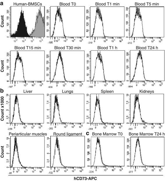
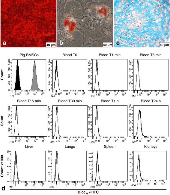
Methods: BMSCs were directly injected in the pig FH, and then the biodistribution of grafted cells was detected by quantitative real-time polymerase chain reaction, cytometry, or a combination of classic histology analysis and in situ hybridization (ISH). BMSC efficacy on bone regeneration was evaluated by magnetic resonance imaging (MRI) and histology.

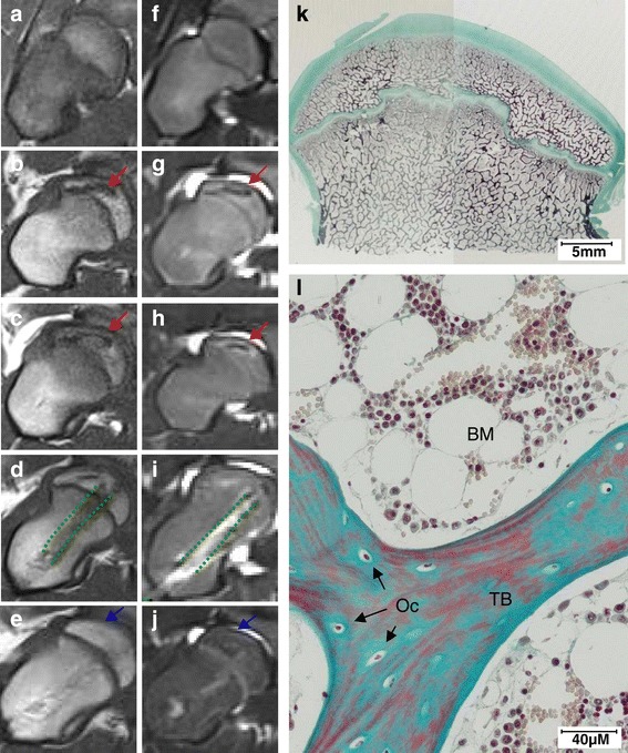
Results: After 30-minute and 24-hour follow-up, grafted cells were detected at the injection site and no BMSCs were detected in filter organs or body fluids. The combination of classic histology analysis and ISH showed a good homogeneity of cell distribution in FH. Local delivery of BMSCs onto a bone scaffold associated with bone formation in vivo confirmed the preferential tropism of BMSCs to the bone tissue as well as their efficacy to form bone. Treatment of a natural pig ONFH by autologous BMSCs indicated a beginning of bone healing as early as 2 weeks with a complete healing after 9 weeks. At this stage, MRI and histological analysis were similar to those of a normal FH.

Conclusions: Intra-osseous injection of BMSCs in FH seems to be a good strategy for ONFH treatment as the safety concerning the biodistribution of BMSCs is ensured. Moreover, the efficacy of BMSCs in natural ONFH seems to indicate that this is a promising approach. Altogether, these results constitute the preclinical data necessary for the setup of a clinical application with expanded BMSCs in the context of advanced therapy medicinal products.


Figures

Figure 1
Figure 1
Biodistribution analysis of injected human bone marrow mesenchymal stromal cells (hBMSCs) in pig femoral head by flow cytometry. hBMSCs were injected in the subchondral area of pig femoral head and analysed at different time point. (a) Blood was collected before (T0) and after injection with a kinetic of 1 minute to 24 hours. (b) Liver, lungs, spleen, and kidneys were analysed either at 30 minutes or 24 hours after injection. Periarticular muscles and round ligament were collected 30 minutes after injection. (c) Bone marrow was collected before (T0) and 24 hours after injection. Dark histogram: unstained hBMSCs; grey histogram: stained CD73-APC hBMSCs; and white histogram: negative control cells of a non-injected pig.
Figure 2
Figure 2
Tracking of injected human cells in pig femoral head by molecular biology. (a) Standard curve of human cells quantified by using human TaqMan Copy Number Reference Assay, RNase P. (b) Conversion of average cycle threshold (Ct) to corresponding number of cells from the straight equation of the standard curve (y = −3.526 × 38.163). (c) Detection (Ct) of injected cells in pig femoral head samples (n = 24) by reverse transcription-quantitative real-time polymerase chain reaction with human RNase P.
Figure 3
Figure 3
Detection of injected cells in pig femoral head (FH) 30 minutes after human bone marrow mesenchymal stromal cell (hBMSCs) injection. (a-e) Masson’s Tri-chrome staining. (a, b) Normal pig FH sections. (c-e) Injected pig FH sections. Arrows indicate the hBMSC area. (e) Localization of sections (c), (d), (h), and (i). (f-i) In situ hybridization of human Alu sequences (Alu-ISH). (f) Negative control tissue of pig FH injected with human cells in omitting Alu probe stained with the hematoxylin only. (g) Negative control tissue of pig FH injected with physiological saline without human cells, with Alu probe and hematoxylin counterstaining. (h, i) Injected pig FH with human cells detected by Alu-probe staining and counterstained with the hematoxylin. The positive nuclei for Alu-probe staining appear in dark brown (arrows). Magnifications: 10× (a, c, f-h), 20× (b, d), 1× (e), and 40× (i).
Figure 4
Figure 4
Biodistribution analysis of injected autologous pig bone marrow mesenchymal stromal cells (BMSCs) in pig femoral head by flow cytometry. Characterization of pig BMSCs in vitro with (a) osteoblastic differentiation with an extensive mineralized matrix of calcium hydroxyapatite crystals stained in red by Alizarin Red S, (b) adipocyte differentiation with adipocytes containing lipid droplets in the cytoplasm stained in red by Oil Red O, and (c) chondrogenic differentiation with chondrocyte-like cells stained by hematoxylin in purple and surrounded by glycosaminoglycan-rich extracellular matrix stained in blue by Alcian Blue. (d) Biodistribution analysis by flow cytometry follow-up during 24 hours (n = 1) of autologous labeled DiOC18-FITC BMSCs injected in subchondral area of pig femoral head. Dark histogram: unstained pig BMSCs; grey histogram: stained DiOC18-FITC pig BMSCs; and white histogram: negative control cells of a non-injected pig. DiOC18, 3, 3′-dioctadecyloxacarbocyanine perchlorate; FITC, fluorescein isothiocyanate.
Figure 5
Figure 5
Histological analysis of in vivo bone formation after 7 weeks of ectopic implantation into immunodeficient mice. Scaffolds (a, b) without pig bone marrow mesenchymal stromal cells (pBMSCs) (a) Magnifications: 4×, (b) Magnifications: 20×. (c, d) with 300,000 pBMSCs directly loaded onto the bone scaffold during the surgery. Cells from three different pigs were tested. (c) Magnifications: 4×, (d) Magnifications: 20×. Decalcified implants (n = 6 per condition) were embedded in paraffin and stained with Masson’s Tri-chrome (blue/green = collagen and non-mineralized bone; red = mineralized bone scaffold; purple = nuclei; and pink = cytoplasm). Dotted lines correspond to the areas of new bone formation. NB, new bone; Sc, scaffold ; FT, fibrous tissue.
Figure 6
Figure 6
Cell therapy of natural femoral head osteonecrosis (nONFH) in the pig with injection of 140 × 106 autologous pig bone marrow mesenchymal stromal cells (pBMSCs) (n = 1) in femoral head (FH). (a) to (j) Coronal magnetic resonance imaging (MRI) analysis: red arrows indicate necrotic area, green dashed lines indicate drilling rearrangement, and blue arrows indicate repair area. (a) to (e) T1-weighted image. (f) to (j) T2-FS (fat saturation)-weighted image. (a) and (f) Normal pig FH (negative control). (b) and (g) Diagnosis of nONFH in pig FH. (c) and (h) Confirmation of nONFH in pig FH 1 month after MRI diagnosis. (d) and (i) Pig FH with nONFH 2 weeks after injection of autologous pBMSCs. (e) and (j) Pig FH with nONFH 9 weeks after injection of autologous pBMSCs. (k) and (l) Histological analysis of pig FH with nONFH 9 weeks after injection of autologous pBMSCs stained with Masson’s Tri-chrome (blue/green = collagen and non-mineralized bone; red = mineralized bone; purple = nuclei). Magnifications: 1× (k) and 20× (l). BM, bone marrow; Oc, osteocytes; TB, trabecular bone.

References

    1. Levasseur R. Mechanisms of osteonecrosis. Joint Bone Spine. 2008;75:639–42. doi: 10.1016/j.jbspin.2008.10.001. - DOI - PubMed
    1. Hungerford DS, Jones LC. Asymptomatic osteonecrosis: Should it be treated? Clin Orthop Relat Res. 2004;429:124–30. doi: 10.1097/01.blo.0000150275.98701.4e. - DOI - PubMed
    1. Hernigou P, Beaujean F. Treatment of osteonecrosis with autologous bone marrow grafting. Clin Orthop Relat Res. 2002;405:14–23. doi: 10.1097/00003086-200212000-00003. - DOI - PubMed
    1. Gangji V, De Maertelaer V, Hauzeur JP. Autologous bone marrow cell implantation in the treatment of non-traumatic osteonecrosis of the femoral head: five year follow-up of a prospective controlled study. Bone. 2011;49:1005–9. doi: 10.1016/j.bone.2011.07.032. - DOI - PubMed
    1. Wang X, Wang Y, Gou W, Lu Q, Peng J, Lu S. Role of mesenchymal stem cells in bone regeneration and fracture repair: A review. Int Orthop. 2013;37:2491–8. doi: 10.1007/s00264-013-2059-2. - DOI - PMC - PubMed

Publication types

MeSH terms

LinkOut - more resources