. 2015 Mar 17;6(1):32.
doi: 10.1186/s13287-015-0013-5.

Characterization of menstrual stem cells: angiogenic effect, migration and hematopoietic stem cell support in comparison with bone marrow mesenchymal stem cells

Affiliations

Characterization of menstrual stem cells: angiogenic effect, migration and hematopoietic stem cell support in comparison with bone marrow mesenchymal stem cells

Francisca Alcayaga-Miranda et al. Stem Cell Res Ther. .

Abstract

Introduction: Stem cells isolated from menstrual fluid (MenSCs) exhibit mesenchymal stem cell (MSCs)-like properties including multi-lineage differentiation capacity. Besides, menstrual fluid has important advantages over other sources for the isolation of MSCs, including ease of access and repeated sampling in a noninvasive manner. Such attributes allow the rapid culture of MenSCs in numbers that are sufficient for therapeutical doses, at lower cell passages.

Methods: In this study, we advance the characterization of MenSC populations in comparison to bone marrow derived mesenchymal stem cells (BM-MSCs) with regards to proliferation, lineage differentiation, migration potential, secretion profile and angiogenic properties in vitro and in a matrigel plug assay in mice. We additionally tested their ability to support hematopoietic stem cell (HSC) expansion in vitro.

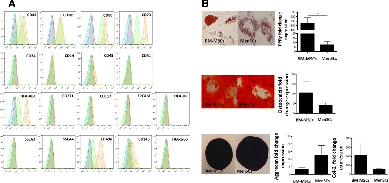
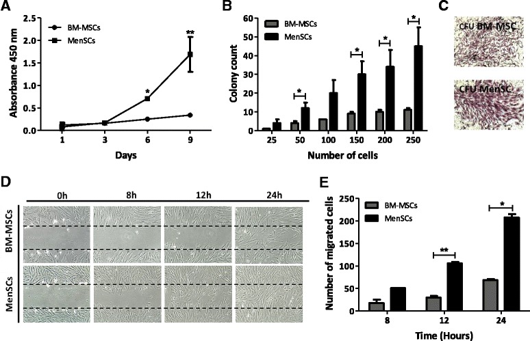
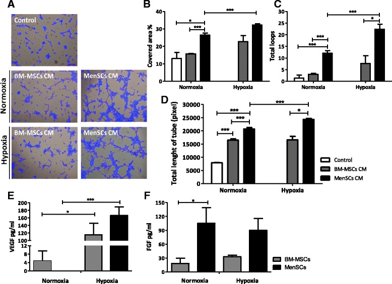
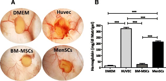
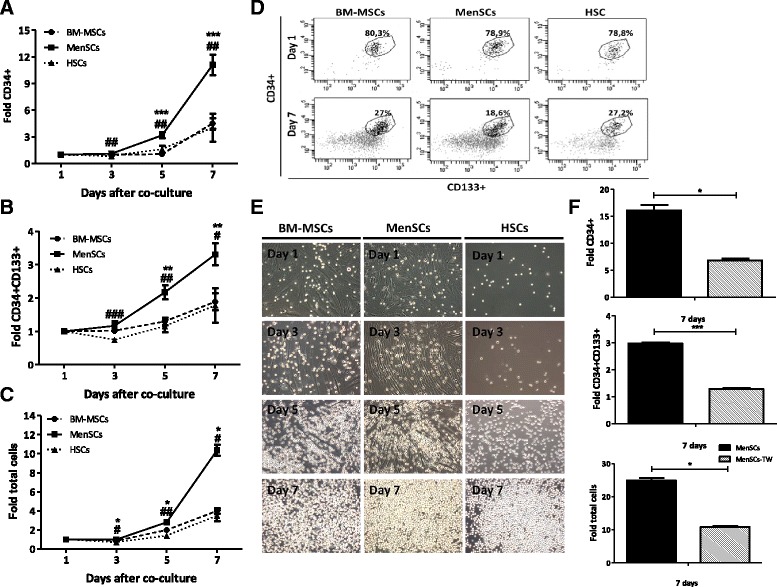
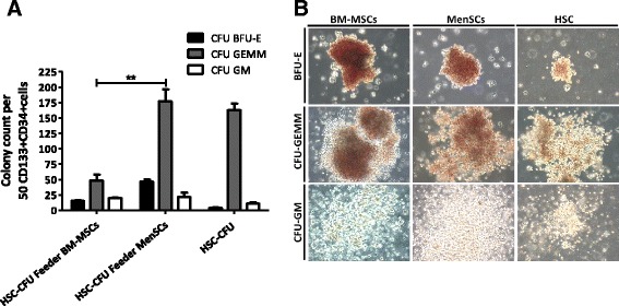
Results: The phenotypic analysis of MenSCs revealed a profile largely similar to the BM-MSCs with the exception of a higher expression of the adhesion molecule CD49a (alpha1-integrin). Furthermore, the fibroblast colony forming units (CFU-F) from MenSCs yielded a 2 to 4 fold higher frequency of progenitors and their in vitro migration capacity was superior to BM-MSCs. In addition, MenSCs evidenced a superior paracrine response to hypoxic conditions as evidenced by the secretion of vascular endothelial growth factor and basic fibroblast growth factor and also improved angiogenic effect of conditioned media on endothelial cells. Furthermore, MenSCs were able to induce angiogenesis in a matrigel plug assay in vivo. Thus, an 8-fold increase in hemoglobin content was observed in implanted plugs containing MenSCs compared to BM-MSCs. Finally, we demonstrated, for the first time, the capacity of MenSCs to support the ex-vivo expansion of HSCs, since higher expansion rates of the CD34+CD133+ population as well as higher numbers of early progenitor (CFU-GEMM) colonies were observed in comparison to the BM source.

Conclusions: We present evidence showing superiority of MenSCs with respect to several functional aspects, in comparison with BM-MSCs. However, the impact of such properties in their use as adult-derived stem cells for regenerative3 medicine remains to be clarified.


Figures

Figure 1
Figure 1
MenSCs show high expression level of mesodermal antigens and multilineage capacities. (A) MenSCs display stem cell-like phenotypic markers. In order to determine the immunophenotype MenSCs and BM-MSCs were stained by conjugated antibodies against mesenchymal, hematopoietic and pluripotential stem cells markers, then analyzed by FACS. MenSCs (orange filled histogram) and BM-MSCs (green filled histogram) displayed positive expression for mesenchymal stromal cells while being negative for other markers, such as CD14, CD34, CD45. Besides, low expression for CD146 and high expression for HLA-ABC and CD49a were observed in MenSCs samples. In addition, FACS analysis showed that both cells do not express pluripotent surface markers, such as SSEA-3, SSEA-4, and TRA-1-60. Isotype-matched controls of MenSCs and BM-MSCs are shown with a blue and red filled histogram, respectively. B) Differentiation potential of BM-MSCs and MenSCs. Adipose differentiation was characterized according to the fold increased level of the peroxysome proliferator-activated receptor (PPAR)-γ and by the formation of lipid droplets that are positive for Oil red O staining. For osteogenesis characterization, the fold increase level of osteocalcin (OC) and positive staining for alizarin red were evaluated. Chondrogenesis was tested by the expression of collagen IIA (col 2A) and aggrecan (Agg) and by positive staining for Safranin O. Values are expressed as mean ± SE of triplicates (*P ≤0.05). Data shown are representative of multiple replicates. BM-MSCs, bone marrow-derived mesenchymal stem cells; FACS, fluorescence-activated cell sorting; MenSCs, menstrual-drived stem cells; SE, standard error.
Figure 2
Figure 2
MenSCs display high proliferation potential, CFU-F capacity, and migration ability. (A) MenSCs show high proliferation. Proliferation was evaluated using cellular mitochondrial dehydrogenase quantification at different time points. MenSCs showed significantly higher proliferation kinetics with respect to BM-MSCs. (B) CFU-F capacity. Serial dilutions of a defined number of cells were cultured and the potential to form CFU was evaluated. MenSCs significantly generated more CFU than BM-MSCs. (C) CFU-F morphology of mesenchymal stem cells. At 21 days of culture, the CFU were stained with crystal violet to visualize the colonies generated. (D-E) Scratch assay. Confluent monolayers of MenSCs and BM-MSCs were mechanically disrupted with a sterile p10 pipet tip. Images were acquired under a phase-contrast microscope. (D) Panels show representative images of BM-MSCs and MenSCs migration post-scratch assay obtained at different time points (magnification 10x). (E) Graph represents statistical analysis of scratch assay. Values are expressed as mean ± SE (*P ≤0.05; **P ≤0.01). Data shown are representative of multiple replicates. BM-MSCs, bone marrow-derived mesenchymal stem cells; CFU-F, fibroblast colony-forming unit; MenSCs, menstrual-drived stem cells; SE, standard error.
Figure 3
Figure 3
Conditioned MenSCs media display significantly higher stimulation of tubule structures formation. HUVEC cells were incubated in MenSCs CM and BM-MSCs CM previously exposed to normoxia (95% O2)/hypoxic conditions (1% O2) or in basal medium, and cultured for six hours with Matrigel. (A) Representative images of the HUVEC tube formation. Panel shows representative images of the capillary network formation on Matrigel assay. Analysis revealed an increase of tubule formation in cells cultured with MenSCs CM, both in normoxia or hypoxia. (B-D) Tubules network quantification. Graphs represent the statistical analysis of the percentage of covered area (B), total loops (C) and total length of tube (D) of tubules network on matrigel angiogenesis assay. HUVEC cultured with MenSCs CM show a statistically significantly higher percentage of covered area, loops and length of tube with respect to the other conditions, both in normoxic and hypoxic conditions. Values shown in the graphs are the mean ± SE (*P ≤0.05; ***P ≤0.001). (E-F) Analysis of soluble factors secreted by MenSCs in vitro. MenSCs and BM-MSCs cells were cultured for 72 hours under normoxic and hypoxic conditions. E) VEGF and F) FGF levels of MenSCs and BM-MSCs CM were assessed by ELISA. Values are expressed as mean ± SE; *,P ≤0.05; ***,P ≤0.001 versus normoxia. Data shown are representative of multiple replicates. BM-MSCs, bone marrow-derived mesenchymal stem cells; CM, conditioned media; FGF, fibroblast growth factor; HUVEC, human umbilical vein endothelial cells; MenSCs, menstrual-drived stem cells; SE, standard error; VEGF, vascular endothelial growth factor.
Figure 4
Figure 4
MenSCs promote angiogenesis in vivo . (A) Matrigel was mixed with cells (3 × 10⁶ cells/plug) or DMEM alone and subcutaneously injected into mice. After 14 days, the matrigel plugs were harvested and photographed. (B) MenSCs have a high hemoglobin content. The hemoglobin content of the plugs from the different groups was measured using Drabkin’s method. The graph represents statistical analysis of the hemoglobin concentration in the matrigel plugs. Values are expressed as mean ± SE (***P ≤0.001). MenSC, menstrual-derived stem cells; SE, standard error.
Figure 5
Figure 5
MenSCs efficiently support the proliferation of CD34+CD133+ hematopoietic stem cells. Cord blood CD34+ cells were immunomagnetically isolated using CD34 beads, then cultured alone or co-cultured at a ratio of 1:4 to 1:5 in different feeder layers from BM-MSCs and MenSCs (contact conditions) or separated by a transwell (TW) system (non-contact conditions). Total number of cells, the expression of CD34+CD133+ hematopoietic stem cells (HSCs) and CD34+ hematopoietic progenitors cells (HPCs) were evaluated at different time points. (A) In vitro expansion of CD34+ hematopoietic progenitor cells. Starting from three days post-culture, a statistically significant difference in favor of HSCs co-cultured with MenSCs was detected. (B) In vitro expansion of CD34+CD133+ HSCs. The in vitro expansion of HSCs showed a 3.31-fold increase (±0.33) with respect to the initial cell numbers when they were co-cultured on the MenSCs-feeder layer. (C) In vitro expansion of total hematopoietic cell numbers. The graph represents the increase of the total cell number obtained in the co-culture on the MenSCs-feeder compared to the BM-MSCs-feeder or alone. Data represent mean ± SE (*P ≤0.05 compared to BM-MSCs; #P ≤0.05 compared to HSCs). (D) Representative FACS analysis seven days post-culture (Dot plot). Percentage of the HSCs at day 1 and day 7. (E) Representative morphology of total hematopoietic cells in co-culture with BM-MSCs and MenSCs. The MenSCs-feeder layer showed a higher capacity to increase the proliferation of HSCs compared to BM-MSCs. Original magnification x10. (F) Analysis of the effect of non-contact expansion conditions. The ex vivo expansion of CD34+ cells, CD34+CD133+ cells, and total cells decreased under the non-contact condition in comparison with the cell contact cultures. Values are expressed as mean ± SE (*P ≤0.05; ***P ≤0.001). Data shown are representative of multiple replicates. BM-MSCs, bone marrow-derived mesenchymal stem cells; MenSCs, menstrual-drived stem cells; SE, standard error.
Figure 6
Figure 6
Ex vivo expansion of CD34 + CD133+ cells on MenSCs-feeder shows high potential of differentiation toward hematopoietic cells. In order to evaluate the progenitors of the hematopoietic lineage, CFU assay was performed by plating a single cell suspension in a semi-solid MethoCult™ media. At 14 days post-culture, colony types derived from the initial equivalent of 50 cord blood HSC was classified and counted according to morphological criteria. (A) Total number of CFU-GEMM, CFU-GM, and BFU-E. Expansion of HSCs on the MenSCs-feeder have a higher capacity to generate CFU-GEMM compared to the other culture conditions. Values are expressed as mean ± SE. (**P ≤0.01). (B) Phase-contrast microscopy of representative CFU-GEMM, CFU-GM and BFU-E colonies. Images are representative of each condition. Original magnification x10. Data shown are representative of multiple replicates. BFU-E, burst-forming unit-erythroid; CFU-GEMM, colony-forming unit-granulocyte, erythroid, macrophage and megakaryocyte; CFU-GM, colony-forming unit-granulocyte and macrophage; HSCs, hematopoietic stem cells; SE, standard error.

Similar articles

Cited by

References

    1. Akimoto K, Kimura K, Nagano M, Takano S, To’a Salazar G, Yamashita T, et al. Umbilical cord blood-derived mesenchymal stem cells inhibit, but adipose tissue-derived mesenchymal stem cells promote, glioblastoma multiforme proliferation. Stem Cells Dev. 2013;22:1370–86. doi: 10.1089/scd.2012.0486. - DOI - PMC - PubMed
    1. Meng X, Ichim TE, Zhong J, Rogers A, Yin Z, Jackson J, et al. Endometrial regenerative cells: a novel stem cell population. J Transl Med. 2007;5:57. doi: 10.1186/1479-5876-5-57. - DOI - PMC - PubMed
    1. Cui CH, Uyama T, Miyado K, Terai M, Kyo S, Kiyono T, et al. Menstrual blood-derived cells confer human dystrophin expression in the murine model of Duchenne muscular dystrophy via cell fusion and myogenic transdifferentiation. Mol Biol Cell. 2007;18:1586–94. doi: 10.1091/mbc.E06-09-0872. - DOI - PMC - PubMed
    1. Hida N, Nishiyama N, Miyoshi S, Kira S, Segawa K, Uyama T, et al. Novel cardiac precursor-like cells from human menstrual blood-derived mesenchymal cells. Stem Cells. 2008;26:1695–704. doi: 10.1634/stemcells.2007-0826. - DOI - PubMed
    1. Patel AN, Park E, Kuzman M, Benetti F, Silva FJ, Allickson JG. Multipotent menstrual blood stromal stem cells: isolation, characterization, and differentiation. Cell Transplant. 2008;17:303–11. doi: 10.3727/096368908784153922. - DOI - PubMed

Publication types

MeSH terms