Comparative Study
. 2015 Feb 13:14:19.
doi: 10.1186/s12934-015-0202-z.

Systematic comparison of single-chain Fv antibody-fusion toxin constructs containing Pseudomonas Exotoxin A or saporin produced in different microbial expression systems

Affiliations
Comparative Study

Systematic comparison of single-chain Fv antibody-fusion toxin constructs containing Pseudomonas Exotoxin A or saporin produced in different microbial expression systems

Pietro Della Cristina et al. Microb Cell Fact. .

Abstract

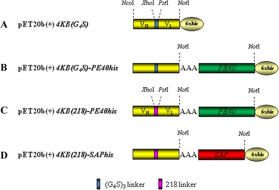
Background: Antibodies raised against selected antigens over-expressed at the cell surface of malignant cells have been chemically conjugated to protein toxin domains to obtain immunotoxins (ITs) able to selectively kill cancer cells. Since latest generation immunotoxins are composed of a toxic domain genetically fused to antibody fragment(s) which confer on the IT target selective specificity, we rescued from the hydridoma 4KB128, a recombinant single-chain variable fragment (scFv) targeting CD22, a marker antigen expressed by B-lineage leukaemias and lymphomas. We constructed several ITs using two enzymatic toxins both able to block protein translation, one of bacterial origin (a truncated version of Pseudomonas exotoxin A, PE40) endowed with EF-2 ADP-ribosylation activity, the other being the plant ribosome-inactivating protein saporin, able to specifically depurinate 23/26/28S ribosomal RNA. PE40 was selected because it has been widely used for the construction of recombinant ITs that have already undergone evaluation in clinical trials. Saporin has also been evaluated clinically and has recently been expressed successfully at high levels in a Pichia pastoris expression system. The aim of the present study was to evaluate optimal microbial expression of various IT formats.

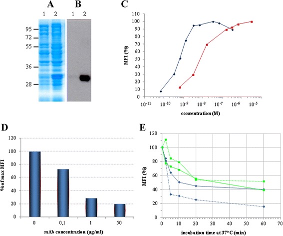
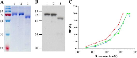
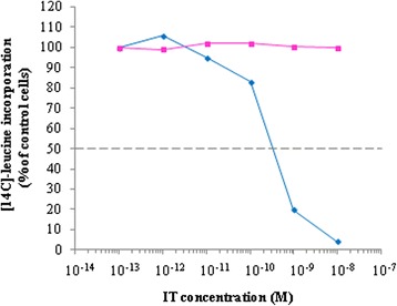
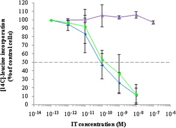
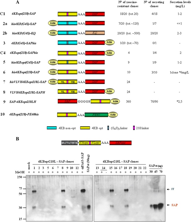
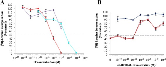
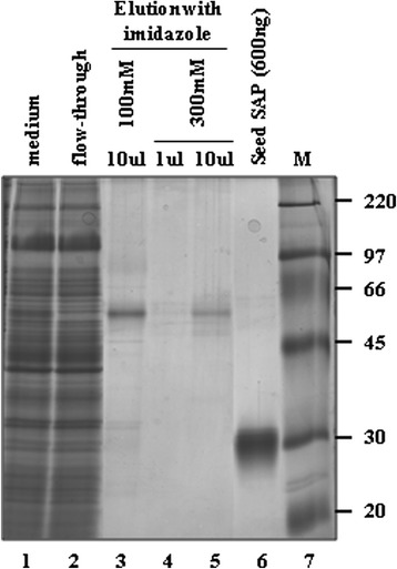
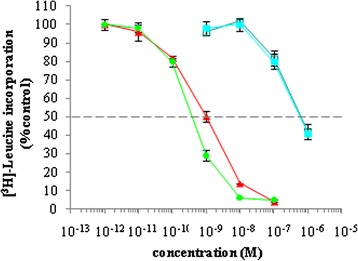
Results: An anti-CD22 scFv termed 4KB was obtained which showed the expected binding activity which was also internalized by CD22+ target cells and was also competed for by the parental monoclonal CD22 antibody. Several fusion constructs were designed and expressed either in E. coli or in Pichia pastoris and the resulting fusion proteins affinity-purified. Protein synthesis inhibition assays were performed on CD22+ human Daudi cells and showed that the selected ITs were active, having IC50 values (concentration inhibiting protein synthesis by 50% relative to controls) in the nanomolar range.

Conclusions: We undertook a systematic comparison between the performance of the different fusion constructs, with respect to yields in E. coli or P. pastoris expression systems and also with regard to each constructs specific killing efficacy. Our results confirm that E. coli is the system of choice for the expression of recombinant fusion toxins of bacterial origin whereas we further demonstrate that saporin-based ITs are best expressed and recovered from P. pastoris cultures after yeast codon-usage optimization.


Figures

Figure 1
Figure 1
Expression & characterization of the 4KB scFv. Total lysate of non-induced (lane 1) and IPTG-induced (lane 2) E. coli BL21(DE3) pLys transformed with pET20b(+)4KBscFv were loaded and the expression of the recombinant protein was detected by (A) Coomassie blue staining or (B) Western blot analysis with anti-His antibody. (C) The binding activity of 4KB scFv (red squares) was compared with that of 4KB128 mAb (blue diamonds) by flow-cytometric analysis on Daudi cells incubated at 4°C using increasing amounts of purified 4KB128 mAb or 4KB scFv. (D) The binding of 4KB scFv (50 μg/ml) on Daudi cells is competitively inhibited by increasing concentrations of the parental anti-CD22 mAb pre-incubated with the cells. The scFv-associated fluorescence without competing mAb pre-incubation is taken as the maximal reference MFI. (E) Internalization and stability of the anti-CD22 mAb compared to 4KB scFv. Ramos (light blue) and Daudi (green) cells were stained at 4°C with 30 μg/ml 4KB scFv (continuous line) or 10 μg/ml mAb (dashed line) and subsequently incubated at 37°C for the indicated times, as described in Methods. Red lines indicate the MFI obtained by staining Daudi cells with the scFv (continuous line) and mAb (dashed line) previously incubated at 37°C for the same time lengths as for the internalization experiment. MFI values are plotted as percentage relative to the fluorescence obtained for samples kept on ice.
Figure 2
Figure 2
Constructs for the expression of toxin-based fusions in E. coli. Schematic representation of 4KBscFv (A), PE (B and C) and saporin (D)-derived constructs. Restriction enzyme sites used for the cloning strategy are also shown (for details, see text under Methods section). Sequence of the 218 linker (218 L) in fuchsia color is: GSTSGSGKPGSGEGSTKG (amino acid one letter code).
Figure 3
Figure 3
Characterization of recombinant ITs expressed in E. coli purified by IMAC. (A) Coomassie staining and (B) Western blot with anti-His antibody of purified 4KB-PE40 in lane 1, 4KB(218)-PE40 in lane 2 and 4KB(218)-SAP in lane 3. (C) Comparison of the binding characteristics of 4KB-PE40 (blue diamonds), 4KB(218)-PE40 (green circles) and 4KB(218)-SAP (red triangles) analyzed by flow-cytometry using Daudi cells incubated at 4°C with increasing concentrations of each IT.
Figure 4
Figure 4
Characterization of 4KB-PE40 IT immunospecificity for CD22 expressed on Daudi cells. The cytotoxic assay was performed incubating Daudi cells for 72 hours with increasing concentrations of 4KB-PE40 in the presence (pink squares) or absence (blue diamonds) of a fixed concentration of the corresponding parental 4KB128 monoclonal antibody. Inhibition of protein synthesis is expressed as percentage of [14C]-leucine incorporation compared to the control samples (untreated cells).
Figure 5
Figure 5
Cytotoxicity of 4KB128-derived rITs for CD22 + Daudi cells. Protein synthesis inhibition assay on Daudi cells exposed for 72 hours to increasing concentrations of 4KB-PE40 (blue diamonds), 4KB(218)-PE40 (green circles) or 4KB(218)-SAP (violet triangles). Protein synthesis inhibition is expressed as a percentage of [14C]-leucine incorporation compared to untreated control cells. Error bars represent standard deviations from the mean of triplicate samples.
Figure 6
Figure 6
4KB-SAP and 4KB-PE40 fusions expressed in Pichia pastoris G115 ( his4 ). (A) Schematic representation of saporin- (C1-9) or PE-based (10) -4KB128-derived fusion constructs. C1: Pichia-fully optimized Construct 1 expressing clone. (B) Western blot analysis of 4KBopt218L-SAP clones. Secretion yields were estimated at 1-2 mg/L and compared to induced mock or positive control anti-PA63scFv-SAP expresser clones.
Figure 7
Figure 7
Cytotoxicity of 4KB128-SAP (C1) produced in P. pastoris for CD22 + Daudi cells. Daudi cells were exposed for 72 hours to increasing concentrations of 4KBscFv-SAP (red triangles), seed SAP (light blue squares) or mock supernatant (violet circles) (A). Inhibition of protein synthesis is expressed as percentage of [3H]-leucine incorporation compared to untreated control cells. Error bars represent standard deviations from the mean of triplicate samples. (B) A competitive inhibition assay was performed by incubating Daudi cells for 72 hours with of 4KB128scFv-SAP at 10−8 M in the presence of increasing concentrations of 4KB128 parental monoclonal antibody (filled and open red circles refer to two different batches of 4KB128 MAb). Inhibition of protein synthesis is expressed as percentage of [3H]-leucine incorporation compared to untreated control cells. Error bars represent standard deviations from the means of triplicate samples. 4KB128 antibody used alone over the full concentration range was not cytotoxic.
Figure 8
Figure 8
Characterization of 4KB128-SAP (C4) produced in P. pastoris and purified by IMAC. Silver staining of purified 4KB128-SAP (C4). MW markers are shown in the far right lane.
Figure 9
Figure 9
Protein synthesis inhibition in Daudi cells exposed for 72 hours to increasing concentrations of 4KB-PE40 produced in E. coli (green circles), C4 (4KBopt218L-SAPHis6) (red triangles), rSAP (open blue squares), seed SAP (solid blue squares). Inhibition of protein synthesis is expressed as percentage of [3H]-leucine incorporation compared to untreated control cells. Error bars represent standard deviations from mean of triplicate samples.
Figure 10
Figure 10
Expression of 4KB218Lopt-PE40 in P. pastoris . A sample from a 72-hour medium scale induction of a GS115 clone expressing 4KB218Lopt-PE40 was analyzed by Western blotting with anti-PE serum. Concentrated medium of the induced culture was loaded in lane 1; 20 ng of recombinant PE40 expressed in E. coli were loaded as a control in lane 2.
Figure 11
Figure 11
Cleavage pattern assessment of secreted PE. (A) Western blot analysis of native PE fragments derived from PE cleaved under different conditions. CTR (control): native PE incubated with PBS; αA BMMY: native PE incubated with BMMY after 48 h of induction of the GS115 mock transformant pPICZαA (αA) clone; αA BMMY PMSF: as αA BMMY but PE was incubated in addition + 1 mM PMSF BMMY: induction medium only. (B) In silico study of identifiable furin-like cleavage sites in the native PE sequence.

Similar articles

Cited by

References

    1. Strebhardt K, Ullrich A. Paul Ehrlich’s magic bullet concept: 100 years of progress. Nat Rev Cancer. 2008;8:473–480. doi: 10.1038/nrc2394. - DOI - PubMed
    1. Vago R, Ippoliti R; Fabbrini, M. S. Current status & Biomedical applications of Ribosome-inactivating proteins. In Antitumor Potential and other Emerging Medicinal Properties of Natural Compounds. Edited by Ng EFFTB: Springer; 2013: 145–179.
    1. Kreitman RJ. Recombinant immunotoxins containing truncated bacterial toxins for the treatment of hematologic malignancies. BioDrugs. 2009;23:1–13. doi: 10.2165/00063030-200923010-00001. - DOI - PMC - PubMed
    1. de Virgilio M, Lombardi A, Caliandro R, Fabbrini MS. Ribosome-inactivating proteins: from plant defense to tumor attack. Toxins (Basel) 2010;2:2699–2737. doi: 10.3390/toxins2112699. - DOI - PMC - PubMed
    1. Fracasso G, Stirpe F, Colombatti M. Plant toxic proteins, ribosome - inactivating protein - containing conjugates for therapeutic use. Toxic Plant Proteins, Plant Cell Monographs. 2010;18:225–263. doi: 10.1007/978-3-642-12176-0_12. - DOI

Publication types

MeSH terms