Jugulotympanic paraganglioma: a rare cause of vertigo
- PMID: 25889842
- PMCID: PMC4407680
- DOI: 10.12659/AJCR.893366
Jugulotympanic paraganglioma: a rare cause of vertigo
Abstract
Background: Jugulotympanic paraganglioma generally presents in the 5th or 6th decades of life with tinnitus and hearing loss. In this manuscript, we present a rare case of jugulotympanic paraganglioma presenting in the 9th decade with vertigo as the most bothersome symptom.
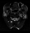
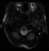
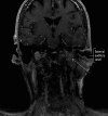
Case report: An 83-year-old woman presented with worsening episodes of dizziness of a few months duration. She also complained of tinnitus and hearing loss, more severe on the left side. Examination revealed a red bulging left-sided tympanic membrane, conductive hearing loss, and a bruit at the base of the skull. Dix-Hallpike test was negative. CT head and MRI brain revealed findings consistent with a large left-sided jugulotympanic paraganglioma, which was found to be hormonally inactive on laboratory tests. The patient underwent treatment with radiotherapy, which resulted in partial improvement of symptoms.
Conclusions: Jugulotympanic paraganglioma may manifest in the elderly with the chief complaint of intermittent vertigo, as in our case. A red bulging mass on otoscopy raises the suspicion, necessitating further investigations, including CT and MRI.
Figures


References
-
- Wong BJ, Roos DE, Borg MF. Glomus jugulare tumours: a 15 year radiotherapy experience in South Australia. J Clin Neurosci Off J Neurosurg Soc Australas. 2014;21(3):456–61. - PubMed
-
- Van den Berg R. Imaging and management of head and neck paragangliomas. Eur Radiol. 2005;15(7):1310–18. - PubMed
-
- Suh JD, Balaker AE, Suh BD, Blackwell KE. Glomus jugulare. Ear Nose Throat J. 2011;90(1):14. - PubMed
Publication types
MeSH terms
LinkOut - more resources
Full Text Sources
Medical

