Comparative Study
. 2015 Mar 24;16(1):43.
doi: 10.1186/s12931-015-0199-1.

Anti-inflammatory effects of novel curcumin analogs in experimental acute lung injury

Affiliations
Comparative Study

Anti-inflammatory effects of novel curcumin analogs in experimental acute lung injury

Yali Zhang et al. Respir Res. .

Abstract

Background: Acute lung injury (ALI) and its most severe form acute respiratory distress syndrome (ARDS) have been the leading cause of morbidity and mortality in intensive care units (ICU). Currently, there is no effective pharmacological treatment for acute lung injury. Curcumin, extracted from turmeric, exhibits broad anti-inflammatory properties through down-regulating inflammatory cytokines. However, the instability of curcumin limits its clinical application.

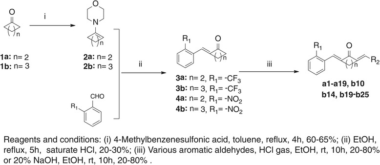
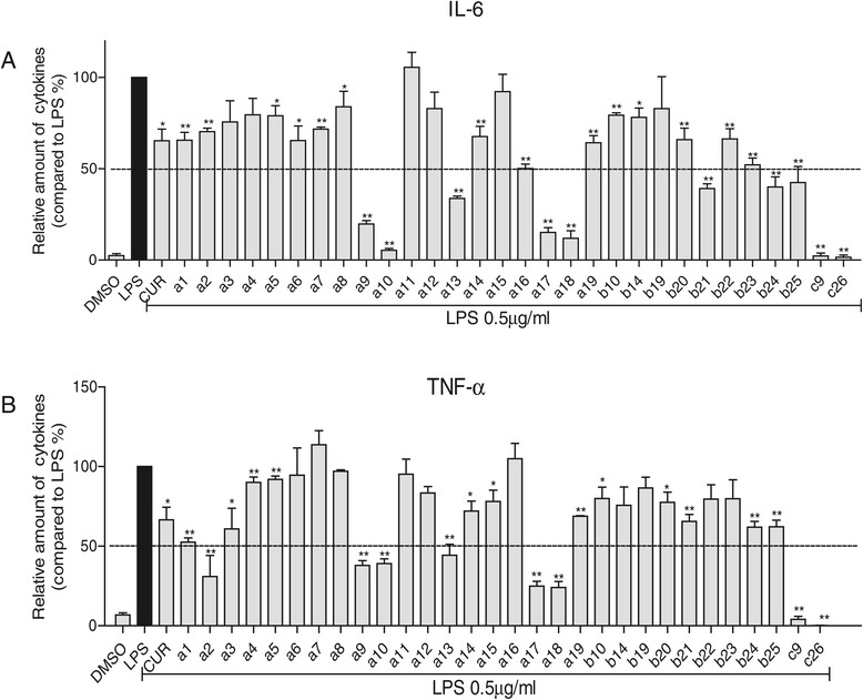
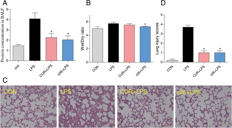
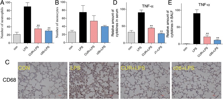
Methods: A series of new curcumin analogs were synthesized and screened for their inhibitory effects on the production of TNF-α and IL-6 in mouse peritoneal macrophages by ELISA. The evaluation of stability and mechanism of active compounds was determined using UV-assay and Western Blot, respectively. In vivo, SD rats were pretreatment with c26 for seven days and then intratracheally injected with LPS to induce ALI. Pulmonary edema, protein concentration in BALF, injury of lung tissue, inflammatory cytokines in serum and BALF, inflammatory cell infiltration, inflammatory cytokines mRNA expression, and MAPKs phosphorylation were analyzed. We also measured the inflammatory gene expression in human pulmonary epithelial cells.

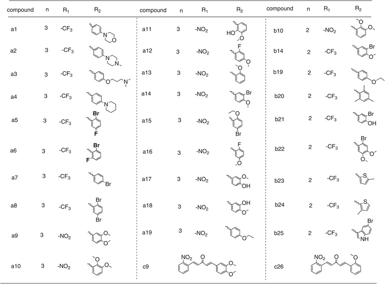
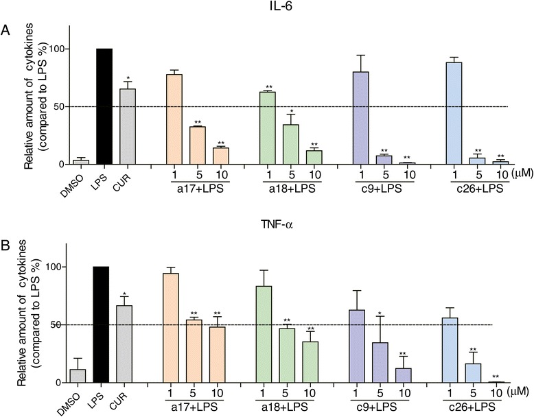
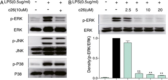
Results: In the study, we synthesized 30 curcumin analogs. The bioscreeening assay showed that most compounds inhibited LPS-induced production of TNF-α and IL-6. The active compounds, a17, a18, c9 and c26, exhibited their anti-inflammatory activity in a dose-dependent manner and exhibited greater stability than curcumin in vitro. Furthermore, the active compound c26 dose-dependently inhibited ERK phosphorylation. In vivo, LPS significantly increased protein concentration and number of inflammatory cells in BALF, pulmonary edema, pathological changes of lung tissue, inflammatory cytokines in serum and BALF, macrophage infiltration, inflammatory gene expression, and MAPKs phosphorylation . However, pretreatment with c26 attenuated the LPS induced increase through ERK pathway in vivo. Meanwhile, compound c26 reduced the LPS-induced inflammatory gene expression in human pulmonary epithelial cells.

Conclusions: These results suggest that the novel curcumin analog c26 has remarkable protective effects on LPS-induced ALI in rat. These effects may be related to its ability to suppress production of inflammatory cytokines through ERK pathway. Compound c26, with improved chemical stability and bioactivity, may have the potential to be further developed into an anti-inflammatory candidate for the prevention and treatment of ALI.


Figures

Scheme 1
Scheme 1
The synthetic pathway of curcumin analogs.
Scheme 2
Scheme 2
The synthetic pathway of curcumin analogs c9 and c26.
Figure 1
Figure 1
The structures of synthesized curcumin analogs.
Figure 2
Figure 2
Production of LPS-induced inflammatory cytokines in mouse peritoneal macrophages after treatment with curcumin derivatives. Macrophages were treated with vehicle or 10 μM curcumin derivatives or 10 μM curcumin for 30 minutes, and the LPS (0.5 μg/mL) was added to incubate for further 24 hours. The culture media was collected and the inflammatory cytokines, IL-6 (A) and TNF-α (B), in media were detected by ELISA and normalized by total protein concentration. The results were presented as the percent of LPS control. Each bar represents mean ± SEM of three independent experiments. Statistical significance relative to LPS group was indicated, *p < 0.05, **p < 0.01.
Figure 3
Figure 3
Active compounds inhibited LPS-induced inflammatory cytokines in a dose-dependent manner. Macrophages were treated with vehicle, 10 μM curcumin or (1, 5, 10 μM) active compounds for 30 minutes, and the LPS (0.5 μg/mL) was added to incubate for further 24 hours. The culture media was collected and the inflammatory cytokines IL-6 (A) and TNF-α (B) in media were detected by ELISA and normalized by total protein concentration. The results were presented as the percent of LPS control. Each bar represents mean ± SEM of three independent experiments. Statistical significance relative to LPS group was indicated, *p < 0.05, **p < 0.01.
Figure 4
Figure 4
UV-visible absorption spectra of curcumin, a17, a18, c9, and c26 in phosphate buffer (pH 7.4), containing 5% DMSO. The compounds stability was described by the curve which is consisted of absorbance at various optical density (250–600 nm) and intervals (0, 5, 10, 15, 20, 25 min). (A) curcumin; (B) a17; (C) a18; (D) c9; (E) c26.
Figure 5
Figure 5
Compound c26 inhibited ERK phosphorylation. (A) Macrophages were cultured with or without c26 for 30 min and stimulated with LPS (0.5 μg/mL) for an additional 20 minutes. Cells were harvested and the total protein was extracted. The protein level of p-ERK, ERK, p-JNK, JNK, p-P38, and P38 was determined by western blot, respectively. (B) Compound c26 dose-dependently decreased ERK phosphorylation. Representative blots of three independent experiments in each study are shown. Statistical significance with regard to LPS was demonstrated, **P <0.01.
Figure 6
Figure 6
Compound c26 attenuated lung pathophysiologic changes in LPS-challenged rats. Effects of c26 on the total protein concentration in (A) BALF and (B) the lung W/D ratio of LPS-induced ALI in rats. Statistical significance relative to LPS group was indicated, *p < 0.05. (C) Compound c26 attenuated LPS-induced histopathological change in lung tissue (H&E staining). (D) The histogram of lung injury scores.
Figure 7
Figure 7
Compound c26 attenuated lung inflammation in LPS-treated rats. Compound c26 reduced LPS-induced number of (A) neutrophils and (B) monocytes in BALF, (C) Compound c26 reduced LPS-induced macrophages infiltration in lung tissue (CD68 immunostaining). Effect of c26 inhibited the inflammatory cytokine TNF-α expression in (D) serum and (E) BALF. Statistical significance relative to LPS group was indicated, *p < 0.05, **p < 0.01.
Figure 8
Figure 8
Effects of c26 inhibited the LPS-induced inflammatory gene expression and inflammatory pathway in lung tissue. (A) TNF-α, (B) IL-6, (C) IL-1β, and (D) COX-2 mRNA expression. (E) The phosphorylation of MAPKs. Statistical significance relative to LPS group was indicated, *p < 0.05, **p < 0.01.
Figure 9
Figure 9
Compound c26 reduced the LPS-induced inflammatory genes expression in human lung epithelial cells. (A) TNF-α, (B) IL-6, (C) IL-1β, (D) COX-2. The results were presented as the percent of LPS control. Each bar represents mean ± SEM of three independent experiments. Statistical significance relative to LPS group was indicated, *p < 0.05, *p < 0.01.

References

    1. Johnson ER, Matthay MA. Acute lung injury: epidemiology, pathogenesis, and treatment. J Aerosol Med Pulm Drug Deliv. 2010;23:243–52. doi: 10.1089/jamp.2009.0775. - DOI - PMC - PubMed
    1. Wheeler AP, Bernard GR. Acute lung injury and the acute respiratory distress syndrome: a clinical review. Lancet. 2007;369:1553–64. doi: 10.1016/S0140-6736(07)60604-7. - DOI - PubMed
    1. Levitt JE, Matthay MA. Clinical review: early treatment of acute lung injury-paradigm shift toward prevention and treatment prior to respiratory failure. Crit Care. 2012;16:223. doi: 10.1186/cc11144. - DOI - PMC - PubMed
    1. Goodman RB, Pugin J, Lee JS, Matthay MA. Cytokine-mediated inflammation in acute lung injury. Cytokine Growth Factor Rev. 2003;14:523–35. doi: 10.1016/S1359-6101(03)00059-5. - DOI - PubMed
    1. Grommes J, Soehnlein O. Contribution of neutrophils to acute lung injury. Mol Med. 2011;17:293. doi: 10.2119/molmed.2010.00138. - DOI - PMC - PubMed

Publication types

MeSH terms