. 2015 Apr 2:12:63.
doi: 10.1186/s12974-015-0282-z.

Insulin improves memory and reduces chronic neuroinflammation in the hippocampus of young but not aged brains

Affiliations

Insulin improves memory and reduces chronic neuroinflammation in the hippocampus of young but not aged brains

Linda Adzovic et al. J Neuroinflammation. .

Erratum in

Abstract

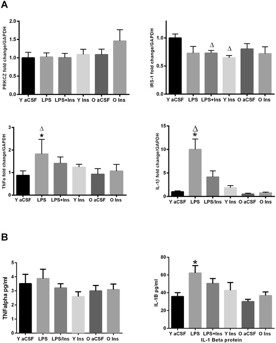
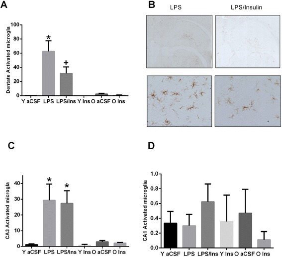
The role of insulin in the brain is still not completely understood. In the periphery, insulin can decrease inflammation induced by lipopolysaccharide (LPS); however, whether insulin can reduce inflammation within the brain is unknown. Experiments administrating intranasal insulin to young and aged adults have shown that insulin improves memory. In our animal model of chronic neuroinflammation, we administered insulin and/or LPS directly into the brain via the fourth ventricle for 4 weeks in young rats; we then analyzed their spatial memory and neuroinflammatory response. Additionally, we administered insulin or artificial cerebral spinal fluid (aCSF), in the same manner, to aged rats and then analyzed their spatial memory and neuroinflammatory response. Response to chronic neuroinflammation in young rats was analyzed in the presence or absence of insulin supplementation. Here, we show for the first time that insulin infused (i.c.v.) to young rats significantly attenuated the effects of LPS by decreasing the expression of neuroinflammatory markers in the hippocampus and by improving performance in the Morris water pool task. In young rats, insulin infusion alone significantly improved their performance as compared to all other groups. Unexpectedly, in aged rats, the responsiveness to insulin was completely absent, that is, spatial memory was still impaired suggesting that an age-dependent insulin resistance may contribute to the cognitive impairment observed in neurodegenerative diseases. Our data suggest a novel therapeutic effect of insulin on neuroinflammation in the young but not the aged brain.


Figures

Figure 1
Figure 1
Morris water maze performance. (A) Latency: in the young groups, the performance was significantly impaired by LPS infusion; performance recovered when insulin was infused with LPS (see text). Surprisingly, the young rats receiving only insulin performed better compared to all other groups. Conversely, in the aged rats, insulin did not improve performance as compared to the controls. (B) Velocity: in the young rats, insulin significantly improved performance versus LPS and LPS/insulin groups (P < 0.05). (C) Distance: the distance to find the platform was significantly smaller in the insulin group (P < 0.05) over the 4 days while it was greater in the LPS group. Insulin/LPS was significant versus LPS (P < 0.05) and versus insulin (P < 0.01). Insulin treatment reduced the distance swam as compared to aCSF (P < 0.05). (D) Probe trial: the performance was impaired by LPS infusion as compared to aCSF and insulin (P < 0.05). (Stars) The aged rats showed no differences, P > 0.05, between aCSF and insulin-infused groups.
Figure 2
Figure 2
RT-PCR and cytokine analyses. (A) Gene expression: PRKCZ gene expression did not differ between groups, contrary to the protein level. IRS-1, top right graph, was decreased in the two young groups receiving insulin as compared to the vehicle group Δ (P < 0.05); in the young rats, IL-1β mRNA was increased in the LPS-infused group, *P < 0.01, versus aCSF and decreased, ΔP < 0.03 versus LPS + insulin. TNFα mRNA was increased by LPS infusion, *P < 0.01, versus aCSF and decreased, ΔP < 0.01, versus LPS + insulin. (B) No significant difference, P > 0.05, in the level of TNFα while IL-1β level was increased, *P < 0.01, as compared to aCSF. The aged rats showed no differences, P > 0.05, between aCSF and insulin-infused groups.
Figure 3
Figure 3
Western blot analyses. The infusion of LPS into the fourth ventricle increased the protein level (A, B) of PKCζ, black square, P < 0.05 versus aCSF. Insulin treatment reduced, black diamond, P < 0.001, PKCζ levels as compared to LPS. (C, D) No significant changes were observed for p-AKT Threonine 308 or Serine 473.
Figure 4
Figure 4
Immunocytochemistry. Photomicrographs and density quantification of MHC II-IR microglial in hippocampus. In the dentate gyrus (A), the density of MHC II-IR was significantly increased by LPS, P < 0.001, and significantly decreased in LPS + insulin rats, P < 0.05. In the CA3 region (C), LPS increased the density of MHC II-IR microglial cells; however, co-infusion of insulin did not decrease the density of MHC II-IR microglia. There were no observed differences in the CA1 region (D) between groups. There were no age-dependent changes in the density of MHC II-IR cells. (B) Scale bar: 400 μm for top row figures; 70 μm for bottom row. *P < 0.05 versus aCSF; + P < 0.05 versus LPS.

References

    1. Akiyama H, Barger S, Barnum S, Bradt B, Bauer J, Cooper NR, et al. Inflammation in Alzheimer’s disease. Neurobiol Aging. 2000;21:383–421. doi: 10.1016/S0197-4580(00)00124-X. - DOI - PMC - PubMed
    1. Lynch AM, Lynch MA. The age-related increase in IL-1 type I receptor in rat hippocampus is coupled with an increase in caspase-3 activation. Eur J Neurosci. 2002;15:1779–88. doi: 10.1046/j.1460-9568.2002.02012.x. - DOI - PubMed
    1. Campuzano O, Castillo-Ruiz MM, Acarin L, Castellano B, Gonzalez B. Increased levels of proinflammatory cytokines in the aged rat brain attenuate injury-induced cytokine response after excitotoxic damage. J Neurosci Res. 2009;87:2484–97. doi: 10.1002/jnr.22074. - DOI - PubMed
    1. Ye SM, Johnson RW. Increased interleukin-6 expression by microglia from brain of aged mice. J Neuroimmunol. 1999;9:139–48. doi: 10.1016/S0165-5728(98)00217-3. - DOI - PubMed
    1. Colton C, Wilcock DM. Assessing activation states in microglia. CNS Neurol Disord Drug Targets. 2010;9:174–91. doi: 10.2174/187152710791012053. - DOI - PubMed

Publication types

MeSH terms