. 2015 Mar 8:12:46.
doi: 10.1186/s12974-015-0256-1.

Anti-MOG antibodies are present in a subgroup of patients with a neuromyelitis optica phenotype

Affiliations

Anti-MOG antibodies are present in a subgroup of patients with a neuromyelitis optica phenotype

Anne-Katrin Pröbstel et al. J Neuroinflammation. .

Abstract

Background: Antibodies against myelin oligodendrocyte glycoprotein (MOG) have been identified in a subgroup of pediatric patients with inflammatory demyelinating disease of the central nervous system (CNS) and in some patients with neuromyelitis optica spectrum disorder (NMOSD). The aim of this study was to examine the frequency, clinical features, and long-term disease course of patients with anti-MOG antibodies in a European cohort of NMO/NMOSD.

Findings: Sera from 48 patients with NMO/NMOSD and 48 patients with relapsing-remitting multiple sclerosis (RR-MS) were tested for anti-aquaporin-4 (AQP4) and anti-MOG antibodies with a cell-based assay. Anti-MOG antibodies were found in 4/17 patients with AQP4-seronegative NMO/NMOSD, but in none of the AQP4-seropositive NMO/NMOSD (n = 31) or RR-MS patients (n = 48). MOG-seropositive patients tended towards younger disease onset with a higher percentage of patients with pediatric (<18 years) disease onset (MOG+, AQP4+, MOG-/AQP4-: 2/4, 3/31, 0/13). MOG-seropositive patients presented more often with positive oligoclonal bands (OCBs) (3/3, 5/29, 1/13) and brain magnetic resonance imaging (MRI) lesions during disease course (2/4, 5/31, 1/13). Notably, the mean time to the second attack affecting a different CNS region was longer in the anti-MOG antibody-positive group (11.3, 3.2, 3.4 years).

Conclusions: MOG-seropositive patients show a diverse clinical phenotype with clinical features resembling both NMO (attacks mainly confined to the spinal cord and optic nerves) and MS with an opticospinal presentation (positive OCBs, brain lesions). Anti-MOG antibodies can serve as a diagnostic and maybe prognostic tool in patients with an AQP4-seronegative NMO phenotype and should be tested in those patients.


Figures
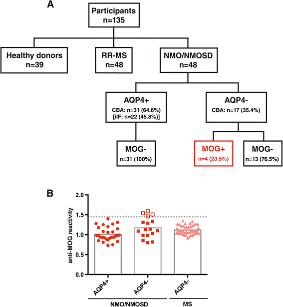
Figure 1
Figure 1
Overview of patient cohort, antibody status, and anti-MOG antibody levels. (A) Patient cohort and antibody status: The study comprised a total of 135 participants including patients with NMO/NMOSD (n = 48), relapsing-remitting multiple sclerosis (RR-MS) (n = 48), and healthy donors (n = 39). Of the NMO/NMOSD patients, 31 were positive for anti-AQP4 antibodies (AQP4+) with the cell-based assay (CBA) while only 22 patients tested positive with indirect immunofluorescence (iIF). All of the AQP4-seropositive patients were negative for anti-MOG antibodies (MOG−). Of the AQP4-seronegative patients (AQP4−), four patients had anti-MOG antibodies (MOG+) (CBA), while 13 patients had no detectable antibodies against either AQP4 or MOG. (B) Anti-MOG antibody levels: Anti-MOG reactivity was analyzed with the CBA and is expressed as the geometric mean channel fluorescence (GMCF) ratio of the MOG-transfected cell line divided by the empty vector-transfected cell line. Values shown represent the (mean) GMCF ratio of one to four experiments. The cutoff used (dotted line) is the mean GMCF ratio of the healthy donor group measured in parallel (n = 39) plus two standard deviations (cutoff = 1.45). Using this cutoff, 4 of the 17 (23.5%) NMO/NMOSD sera were positive for anti-MOG antibodies (empty red squares), all of which were AQP4-seronegative (filled red squares). None of the AQP4-seropositive NMO/NMOSD patients (filled red circles) and none of the RR-MS patients (filled light red triangles) were positive for anti-MOG antibodies.
Figure 2
Figure 2
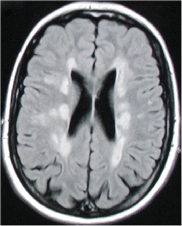
Brain MRI abnormalities in an anti-MOG antibody-positive patient. Axial T2-FLAIR (fluid-attenuated inversion recovery) brain MRI of a MOG-seropositive NMO patient 31 years after disease manifestation with combined optic neuritis and transverse myelitis showed multiple abnormalities fulfilling Barkhof criteria and resembling multiple sclerosis lesions.
Figure 3
Figure 3
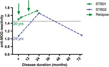
Longitudinal follow-up of MOG-seropositive patients of up to 6 years reveals fluctuating anti-MOG antibody levels. Longitudinal serum samples were available for two of the four anti-MOG antibody-positive patients. Serum samples were taken 30 or 29 years after disease onset over a time course of about 2 or 6 years (22 or 74 months). Anti-MOG reactivity is expressed as the geometric mean channel fluorescence (GMCF) ratio. The cutoff used (dotted line) is the mean GMCF ratio of the healthy donor group measured in parallel (n = 39) plus two standard deviations (cutoff = 1.45). The arrow indicates a clinical relapse. Antibodies against MOG fluctuated over the disease course: An increase of anti-MOG antibodies was associated in one patient (STB01) with an increased relapse frequency, but was independent of disease activity in the other patient (STB02).

References

    1. de Seze J, Stojkovic T, Ferriby D, Gauvrit J-Y, Montagne C, Mounier-Vehier F, et al. Devic’s neuromyelitis optica: clinical, laboratory, MRI and outcome profile. J Neurol Sci. 2002;197:57–61. doi: 10.1016/S0022-510X(02)00043-6. - DOI - PubMed
    1. Wingerchuk DM, Lennon VA, Pittock SJ, Lucchinetti CF, Weinshenker BG. Revised diagnostic criteria for neuromyelitis optica. Neurology. 2006;66:1485–9. doi: 10.1212/01.wnl.0000216139.44259.74. - DOI - PubMed
    1. Wingerchuk DM, Lennon VA, Lucchinetti CF, Pittock SJ, Weinshenker BG. The spectrum of neuromyelitis optica. Lancet Neurol. 2007;6:805–15. doi: 10.1016/S1474-4422(07)70216-8. - DOI - PubMed
    1. Lennon VA, Wingerchuk DM, Kryzer TJ, Pittock SJ, Lucchinetti CF, Fujihara K, et al. A serum autoantibody marker of neuromyelitis optica: distinction from multiple sclerosis. Lancet. 2004;364:2106–12. doi: 10.1016/S0140-6736(04)17551-X. - DOI - PubMed
    1. Bradl M, Misu T, Takahashi T, Watanabe M, Mader S, Reindl M, et al. Neuromyelitis optica: pathogenicity of patient immunoglobulin in vivo. Ann Neurol. 2009;66:630–43. doi: 10.1002/ana.21837. - DOI - PubMed

Publication types

LinkOut - more resources