. 2015 Apr 1:12:61.
doi: 10.1186/s12974-015-0284-x.

The inhibitory effect of mesenchymal stem cell on blood-brain barrier disruption following intracerebral hemorrhage in rats: contribution of TSG-6

Affiliations

The inhibitory effect of mesenchymal stem cell on blood-brain barrier disruption following intracerebral hemorrhage in rats: contribution of TSG-6

Min Chen et al. J Neuroinflammation. .

Abstract

Background: Mesenchymal stem cells (MSCs) are well known having beneficial effects on intracerebral hemorrhage (ICH) in previous studies. The therapeutic mechanisms are mainly to investigate proliferation, differentiation, and immunomodulation. However, few studies have used MSCs to treat blood-brain barrier (BBB) leakage after ICH. The influence of MSCs on the BBB and its related mechanisms were investigated when MSCs were transplanted into rat ICH model in this study.

Methods: Adult male Sprague-Dawley (SD) rats were randomly divided into sham-operated group, PBS-treated (ICH + PBS) group, and MSC-treated (ICH + MSC) group. ICH was induced by injection of IV collagenase into the rats' brains. MSCs were transplanted intravenously into the rats 2 h after ICH induction in MSC-treated group. The following factors were compared: inflammation, apoptosis, behavioral changes, inducible nitric oxide synthase (iNOS), matrix metalloproteinase 9 (MMP-9), peroxynitrite (ONOO(-)), endothelial integrity, brain edema content, BBB leakage, TNF-α stimulated gene/protein 6 (TSG-6), and nuclear factor-κB (NF-κB) signaling pathway.

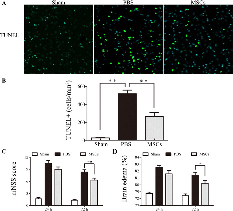
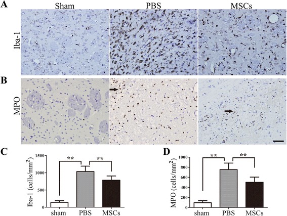
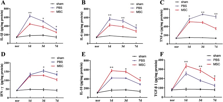
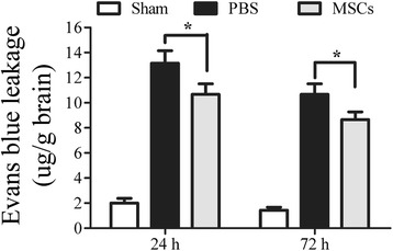
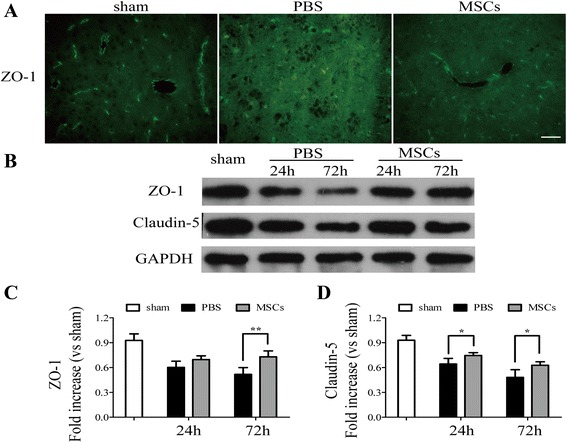
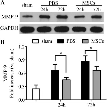
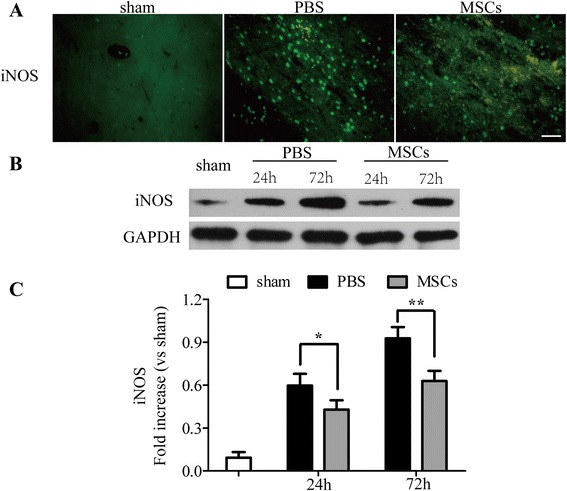
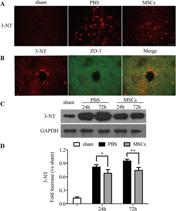
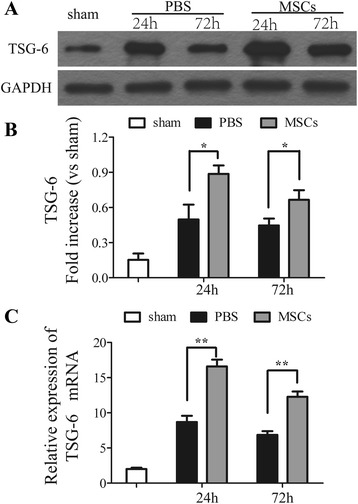
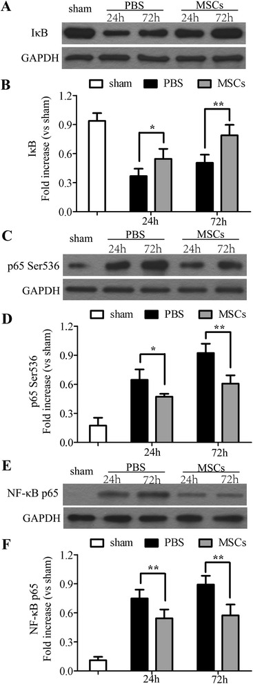
Results: In the ICH + MSC group, MSCs decreased the levels of proinflammatory cytokines and apoptosis, downregulated the density of microglia/macrophages and neutrophil infiltration at the ICH site, reduced the levels of iNOS and MMP-9, attenuated ONOO(-) formation, and increased the levels of zonula occludens-1 (ZO-1) and claudin-5. MSCs also improved the degree of brain edema and BBB leakage. The protective effect of MSCs on the BBB in ICH rats was possibly invoked by increased expression of TSG-6, which may have suppressed activation of the NF-κB signaling pathway. The levels of iNOS and ONOO(-), which played an important role in BBB disruption, decreased due to the inhibitory effects of TSG-6 on the NF-κB signaling pathway.

Conclusions: Our results demonstrated that intravenous transplantation of MSCs decreased the levels of ONOO(-) and degree of BBB leakage and improved neurological recovery in a rat ICH model. This strategy may provide a new insight for future therapies that aim to prevent breakdown of the BBB in patients with ICH and eventually offer therapeutic options for ICH.


Figures

Figure 1
Figure 1
Effect of MSC transplantation on apoptosis, functional recovery, and brain water content. Influence of MSC transplantation on apoptosis, mNSS, and brain water content. Compared with the PBS-treated group, the number of TUNEL-positive cells in the cortical hemorrhagic boundary in the MSC group was significantly decreased at 72 h after ICH (A, B). The mNSS and brain water content were tested 24 and 72 h after ICH. Treatment with MSCs significantly lowered mNSS at 24 and 72 h. The mNSS was differed significantly 72 h after ICH between the PBS- and MSC- treated groups (C). The PBS-treated group had a significantly higher brain water content than the sham-operated control group. MSC treatment reduced brain water content compared with the PBS-treated group 24 and 72 h after ICH. The brain water content was different between the two groups 72 h after ICH (D). n = 6 in each time point per group. Data are presented as the mean ± SD. *P < 0.05; **P < 0.01. Original magnification, × 600. mNSS, modified neurological severity score; MSCs, mesenchymal stem cells; PBS, phosphate-buffered saline; TUNEL, Terminal deoxynucleotidyl transferase-mediated biotinylated-dUTP nick-end labeling.
Figure 2
Figure 2
The influence of MSC on brain inflammatory cell infiltration and microglia numbers. Iba-1+ microglia cells/macrophages and MPO+ neutrophils were identified by immunohistochemistry 72 h after ICH to test the effects of MSC treatment on the number of peripheral infiltrating and brain-resident immune cells. Both the numbers of Iba-1+ microglia cells/macrophages (A, C) and infiltrated MPO+ neutrophils (B, D) were reduced in the MSC-treated group when compared with the PBS-treated group. The sign of arrow indicates the edge of the hematoma. n = 6 per group. Data are presented as the mean ± SD. Bar = 50 μm. **P < 0.01. MPO, myeloperoxidase; MSCs, mesenchymal stem cells; PBS, phosphate-buffered saline.
Figure 3
Figure 3
Influence of MSC treatment on cytokine concentrations. Levels of the proinflammatory cytokines IL-1β (at 1, 3, and 7 days), IL-6 (at 1, 3, and 7 days), TNF-α (at 1, 3, and 7 days), and IFN-γ (at 3 and 7 days) were decreased in the MSC-treated group compared with the PBS-treated group (A-D). Levels of the anti-inflammatory cytokines IL-10 (at 1, 3, and 7 days) and TGF-β1 (at 1, 3, and 7 days) were increased in the MSC-treated group compared with the PBS-treated group (E-F). n = 6 in each time point per group. Data are presented as the mean ± SD. *P < 0.05; **P < 0.01. MSCs, mesenchymal stem cells; PBS, phosphate-buffered saline.
Figure 4
Figure 4
Influence of MSC treatment on blood–brain barrier permeability. The intensity of Evan’s blue determined by spectrofluorometry showed that administration of MSCs reduced BBB leakage when compared with the PBS-treated group 24 and 72 h after ICH. n = 6 in each time point per group. Data are presented as the mean ± SD. *P < 0.05.
Figure 5
Figure 5
Transplantation of MSCs increased the levels of zonula occludens-1 (ZO-1) and claudin-5. Immunofluorescence analysis of ZO-1 and western blotting analysis of ZO-1 and claudin-5. Immunofluorescence analysis of ZO-1 (A) showed that treatment of MSCs increased the levels of tight junction protein compared with the PBS-treated group 72 h after ICH. Western blotting analysis of ZO-1 (B, C) and claudin-5 (B, D) showed similar results in that transplantation of MSCs upregulated the levels of tight junctions compared with the PBS-treated group 24 and 72 h after ICH. n = 6 in each time point per group. Data are presented as the mean ± SD. Bar = 50 μm. *P < 0.05; **P < 0.01. GAPDH, glyceraldehyde 3-phosphate dehydrogenase; MSCs, mesenchymal stem cells; PBS, phosphate-buffered saline.
Figure 6
Figure 6
Transplantation of MSC decreased the levels of matrix metalloproteinase-9 (MMP-9). Western blotting analysis of MMP-9. Treatment with MSCs downregulated the levels of MMP-9 24 and 72 h after ICH when compared with the PBS-treated group (A, B). n = 6 in each time point per group. Data are presented as the mean ± SD. *P < 0.05. GAPDH, glyceraldehyde 3-phosphate dehydrogenase; MMP-9, matrix metalloproteinase 9; MSCs, mesenchymal stem cells; PBS, phosphate-buffered saline.
Figure 7
Figure 7
Transplantation of MSC decreased the levels of inducible nitric oxide synthase (iNOS). Immunofluorescence and western blotting analysis of iNOS. Immunofluorescence analysis of iNOS (A) showed that treatment with MSCs decreased the levels of iNOS compared with the PBS-treated group 72 h after ICH. Western blotting (B, C) analysis of iNOS showed similar results in that transplantation of MSCs downregulated the levels of iNOS compared with the PBS-treated group 24 and 72 h after ICH. n = 6 in each time point per group. Data are presented as the mean ± SD. Bar = 50 μm. *P < 0.05; **P < 0.01. GAPDH, glyceraldehyde 3-phosphate dehydrogenase; iNOS, inducible nitric oxide synthase; MSCs, mesenchymal stem cells; PBS, phosphate-buffered saline.
Figure 8
Figure 8
Transplantation of MSCs decreased the levels of 3-nitrotyrosine (3-NT). Immunofluorescence and western blotting analysis of 3-NT. Immunofluorescence analysis of 3-NT (A) showed that the treatment with MSCs decreased the levels of 3-NT compared with the PBS-treated group 72 h after ICH. Western blotting (C, D) analysis of 3-NT showed the similar results in that transplantation of MSCs downregulated the levels of 3-NT compared with the PBS-treated group 24 and 72 h after ICH. The double labeling of 3-NT and ZO-1 indicated that 3-NT and vascular damage are closely related (B). n = 6 in each time point per group. Data are presented as the mean ± SD. Bar = 50 μm. *P < 0.05; **P < 0.01. GAPDH, glyceraldehyde 3-phosphate dehydrogenase; MSCs, mesenchymal stem cells; PBS, phosphate-buffered saline.
Figure 9
Figure 9
Influence of MSC treatment on TNF-α stimulated gene/protein 6 (TSG-6). Western blotting and real-time PCR analysis of TSG-6. Transplantation of MSCs increased the levels of TSG-6 24 and 72 h after ICH compared with the PBS-treated group (A, B). Similar results were observed at the mRNA levels of TSG-6 (C). n = 6 in each time point per group. Data are presented as the mean ± SD. *P < 0.05; **P < 0.01. GAPDH, glyceraldehyde 3-phosphate dehydrogenase; MSCs, mesenchymal stem cells; PBS, phosphate-buffered saline; TSG-6, TNF-α stimulated gene/protein 6.
Figure 10
Figure 10
Effects of MSC on NF-κB signaling pathway. Treatment with MSCs suppressed activation of the NF-κB signaling pathway 24 and 72 h after ICH. By Western blotting analysis, a basal level of IκB-α (A, B) was detected in the brain tissue from sham-operated rats, whereas in the PBS-treated rats, IκB-α levels were substantially reduced. MSC treatment prevented the degradation of IκB-α in the PBS-treated group. Phosphorylation of Ser536 (C, D) in the cytoplasm and phosphorylation of NF-κB p65 (E, F) levels in nuclear fractions were increased in the PBS-treated group when compared with the sham-operated group. MSC treatment significantly reduced the phosphorylation of p65 on Ser536 and NF-κB p65 levels. n = 6 in each time point per group. Data are presented as the mean ± SD. *P < 0.05; **P < 0.01. GAPDH, glyceraldehyde 3-phosphate dehydrogenase; MSCs, mesenchymal stem cells; NF-κB, nuclear factor-кB; PBS, phosphate-buffered saline.

References

    1. Butcher K, Laidlaw J. Current intracerebral haemorrhage management. J Clin Neurosci. 2003;10:158–67. doi: 10.1016/S0967-5868(02)00324-7. - DOI - PubMed
    1. Xi G, Keep RF, Hoff JT. Erythrocytes and delayed brain edema formation following intracerebral hemorrhage in rats. J Neurosurg. 1998;89:991–6. doi: 10.3171/jns.1998.89.6.0991. - DOI - PubMed
    1. Yang GY, Betz AL, Chenevert TL, Brunberg JA, Hoff JT. Experimental intracerebral hemorrhage: relationship between brain edema, blood flow, and blood–brain barrier permeability in rats. J Neurosurg. 1994;81:93–102. doi: 10.3171/jns.1994.81.1.0093. - DOI - PubMed
    1. Abbott NJ, Patabendige AA, Dolman DE, Yusof SR, Begley DJ. Structure and function of the blood–brain barrier. Neurobiol Dis. 2010;37:13–25. doi: 10.1016/j.nbd.2009.07.030. - DOI - PubMed
    1. Kim KS. Mechanisms of microbial traversal of the blood–brain barrier. Nat Rev Microbiol. 2008;6:625–34. doi: 10.1038/nrmicro1952. - DOI - PMC - PubMed

Publication types

MeSH terms

LinkOut - more resources