. 2015 Feb 15:13:52.
doi: 10.1186/s12957-015-0465-1.

Bone marrow mesenchymal stem cells promote osteosarcoma cell proliferation and invasion

Affiliations

Bone marrow mesenchymal stem cells promote osteosarcoma cell proliferation and invasion

Fu-Xiang Yu et al. World J Surg Oncol. .

Abstract

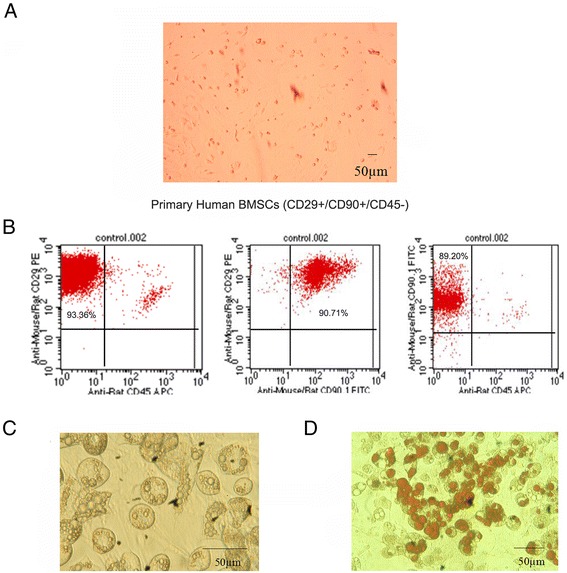
Background: Bone marrow-derived stem cells (BMSCs) are locally adjacent to the tumor tissues and may interact with tumor cells directly. The purpose of this study was to explore the effects of BMSCs on the proliferation and invasion of osteosarcoma cells in vitro and the possible mechanism involved.

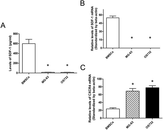
Methods: BMSCs were co-cultured with osteosarcoma cells, and CCK-8 assay was used to measure cell proliferation. The ELISA method was used to determine the concentration of stromal cell-derived factor-1 (SDF-1) in the supernatants. Reverse transcription polymerase chain reaction (RT-PCR) was performed to detect the expression of CXCR4 in osteosarcoma cells and BMSCs. Matrigel invasion assay was performed to measure tumor cell invasion.

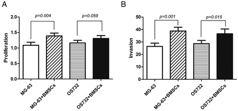
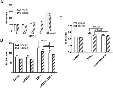
Results: SDF-1 was detected in the supernatants of BMSCs, but not in osteosarcoma cells. Higher CXCR4 mRNA levels were detected in the osteosarcoma cell lines compared to BMSCs. In addition, conditioned medium from BMSCs can promote the proliferation and invasion of osteosarcoma cells, and AMD3100, an antagonist for CXCR4, can significantly downregulate these growth-promoting effects.

Conclusions: BMSCs can promote the proliferation and invasion of osteosarcoma cells, which may involve the SDF-1/CXCR4 axis.


Figures

Figure 1
Figure 1
Identification of bone marrow-derived stem cells. (A) The phenotype of BMSCs isolated from human bone marrow tissue (on the first day; 100× magnification fold). (B) The expression of CD45, CD29, and CD90 in isolated BMSCs. (C) The fat droplets were observed in the cytoplasm of adipocytes at 400× magnification. (D) The fat droplets were stained with Oil red O. Image is magnified 200 ×.
Figure 2
Figure 2
Effect of BMSCs on the proliferation and invasion of osteosarcoma cells. (A) After co-culturing BMSCs with osteosarcoma cells for 72 h, the proliferation of osteosarcoma cells was examined using the CCK-8 assay. The proliferation of MG-63 and OS732 cells co-cultured with BMSCs was significantly increased compared to that of the respective cells cultured alone. (B) BMSCs were co-cultured with osteosarcoma cells for 72 h, and the percentage of osteosarcoma cell invasion was examined under a microscope. The invasion of MG-63 and OS732 cells was significantly greater in the presence of BMSCs compared to control cells.
Figure 3
Figure 3
SDF-1 secretion and SDF-1 and CXCR4 expression. (A) The SDF-1 concentration in the BMSC supernatant as detected by ELISA. BMSCs had significantly higher levels of SDF-1 compared to MG-63 and OS732 cells. (B) The RT-PCR analysis of SDF-1 mRNA expression in osteosarcoma cell lines and BMSCs. (C) The RT-PCR analysis of CXCR4 mRNA expression in osteosarcoma cell lines and BMSCs. (*P < 0.01 compared to the BMSCs).
Figure 4
Figure 4
Effects of SDF-1 on the proliferation of osteosarcoma cell lines. (A) SDF-1 promotes the proliferation of osteosarcoma cells in a dose-dependent manner. (B) AMD3100 partially blocks SDF-1-mediated proliferation. (C) AMD3100 downregulates the effects of BMSCs on osteosarcoma cell line proliferation.

References

    1. Bhowmick NA, Neilson EG, Moses HL. Stromal fibroblasts in cancer initiation and progression. Nature. 2004;432(7015):332–7. doi: 10.1038/nature03096. - DOI - PMC - PubMed
    1. Armstrong T, Packham G, Murphy LB, Bateman AC, Conti JA, Fine DR, et al. Type I collagen promotes the malignant phenotype of pancreatic ductal adenocarcinoma. Clin Cancer Res. 2004;10(21):7427–37. doi: 10.1158/1078-0432.CCR-03-0825. - DOI - PubMed
    1. Bachem MG, Schunemann M, Ramadani M, Siech M, Beger H, Buck A, et al. Pancreatic carcinoma cells induce fibrosis by stimulating proliferation and matrix synthesis of stellate cells. Gastroenterology. 2005;128(4):907–21. doi: 10.1053/j.gastro.2004.12.036. - DOI - PubMed
    1. Miyamoto H, Murakami T, Tsuchida K, Sugino H, Miyake H, Tashiro S. Tumor-stroma interaction of human pancreatic cancer: acquired resistance to anticancer drugs and proliferation regulation is dependent on extracellular matrix proteins. Pancreas. 2004;28(1):38–44. doi: 10.1097/00006676-200401000-00006. - DOI - PubMed
    1. Muerkoster S, Wegehenkel K, Arlt A, Witt M, Sipos B, Kruse ML, et al. Tumor stroma interactions induce chemoresistance in pancreatic ductal carcinoma cells involving increased secretion and paracrine effects of nitric oxide and interleukin-1beta. Cancer Res. 2004;64(4):1331–7. doi: 10.1158/0008-5472.CAN-03-1860. - DOI - PubMed

Publication types

MeSH terms