. 2015 Apr 16;6(1):79.
doi: 10.1186/s13287-015-0073-6.

Umbilical cord mesenchymal stem cells modulate dextran sulfate sodium induced acute colitis in immunodeficient mice

Affiliations

Umbilical cord mesenchymal stem cells modulate dextran sulfate sodium induced acute colitis in immunodeficient mice

Antara Banerjee et al. Stem Cell Res Ther. .

Abstract

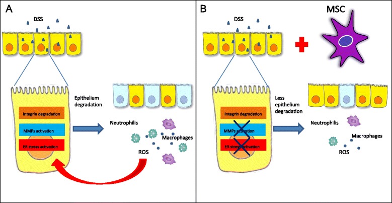
Introduction: Inflammatory bowel diseases (IBD) are complex multi-factorial diseases with increasing incidence worldwide but their treatment is far from satisfactory. Unconventional strategies have consequently been investigated, proposing the use of cells as an effective alternative approach to IBD. In the present study we examined the protective potential of exogenously administered human umbilical cord derived mesenchymal stem cells (UCMSCs) against Dextran Sulfate Sodium (DSS) induced acute colitis in immunodeficient NOD.CB17-Prkdc (scid)/J mice with particular attention to endoplasmic reticulum (ER) stress.

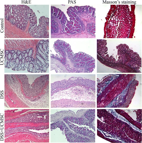
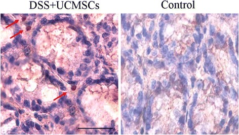
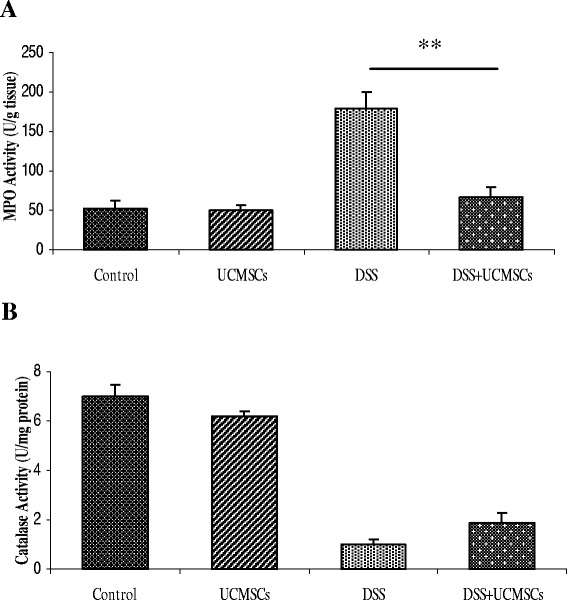
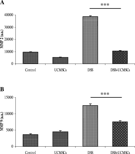
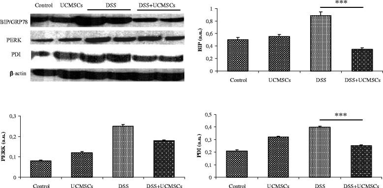
Methods: UCMSCs were injected in NOD.CB17-Prkdc (scid)/J via the tail vein at day 1 and 4 after DSS administration. To verify attenuation of DSS induced damage by UCMSCs, Disease Activity Index (DAI) and body weight changes was monitored daily. Moreover, colon length, histological changes, myeloperoxidase and catalase activities, metalloproteinase (MMP) 2 and 9 expression and endoplasmic reticulum (ER) stress related proteins were evaluated on day 7.

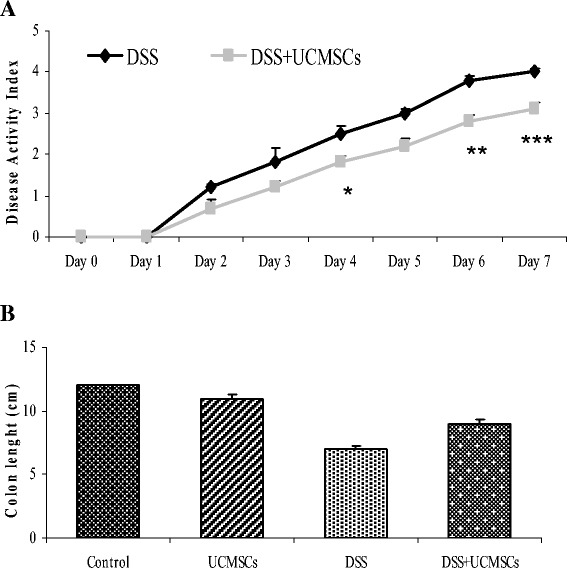
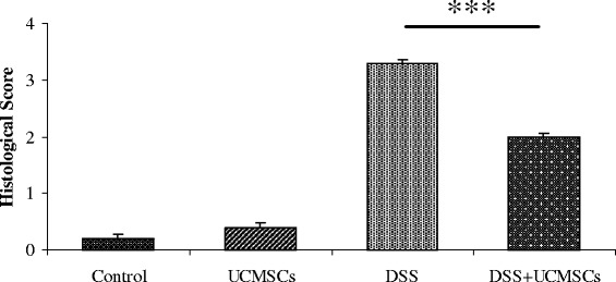
Results: UCMSCs administration to immunodeficient NOD.CB17-Prkdc (scid)/J mice after DSS damage significantly reduced DAI (1.45 ± 0.16 vs 2.08 ± 0.18, p < 0.05), attenuating the presence of bloody stools, weight loss, colon shortening (8.95 ± 0.33 cm vs 6.8 ± 0.20 cm, p < 0.01) and histological score (1.97 ± 0.13 vs 3.27 ± 0.13, p < 0.001). Decrease in neutrophil infiltration was evident from lower MPO levels (78.2 ± 9.7 vs 168.9 ± 18.2 U/g, p < 0.01). DSS treatment enhanced MMP2 and MMP9 activities (>3-fold), which were significantly reduced in mice receiving UCMSCs. Moreover, positive modulation in ER stress related proteins was observed after UCMSCs administration.

Conclusions: Our results demonstrated that UCMSCs are able to prevent DSS-induced colitis in immunodeficient mice. Using these mice we demonstrated that our UCMSCs have a direct preventive effect other than the T-cell immunomodulatory properties which are already known. Moreover we demonstrated a key function of MMPs and ER stress in the establishment of colitis suggesting them to be potential therapeutic targets in IBD treatment.


Figures

Figure 1
Figure 1
Cellular characterization of umbilical cord mesenchymal stem cells. Flow cytometry analysis of umbilical cord mesenchymal stem cells showed a mesenchymal phenotype. Cells were positive for typical mesenchymal markers (CD29, CD73, CD90, CD105 and CD166) while hematopoietic markers (CD44 and c-kit) were weakly or not expressed. Human leukocyte antigen-DR (HLA-DR) was not expressed at all.
Figure 2
Figure 2
Clinical and therapeutic efficacy of exogenous umbilical cord mesenchymal stem cell administration. (A) Disease activity index (DAI). In the umbilical cord mesenchymal stem cells (UCMSCs)-treated group, the DAI score was significantly downregulated on days 4, 6 and 7 of treatment. (B) Colon length. UCMSCs in dextran sulfate sodium (DSS)-treated mice were able to reduce the degree of DSS-induced colon shortening compared with the DSS + phosphate-buffered saline group. Values reported as mean ± standard deviation. *P <0.05, **P <0.01, ***P <0.001.
Figure 3
Figure 3
Therapeutic efficacy of umbilical cord mesenchymal stem cell treatment on the histological colitis score. Administration of umbilical cord mesenchymal stem cells (UCMSCs) in dextran sulfate sodium (DSS)-treated mice significantly improved histological scores. Values reported as mean ± standard deviation. ***P <0.001.
Figure 4
Figure 4
Histological analysis of the distal colon on day 7 of control mice (no dextran sulfate sodium (DSS)), umbilical cord mesenchymal stem cell-treated mice, DSS + phosphate-buffered saline-treated mice and DSS + umbilical cord mesenchymal stem cell-treated mice. Hematoxylin and eosin (H&E) staining showed improvement after umbilical cord mesenchymal stem cell (UCMSCs) treatment because it reduced the extent of the inflamed area, crypt damage, edema of submucosa and infiltration of inflammatory cells. Periodic acid–Schiff (PAS) staining detected goblet cells that displayed a strong purple/magenta color when stained with PAS reagent. There was complete depletion of goblet cells in the crypts of the dextran sulfate sodium (DSS) + phosphate-buffered saline mice. In contrast, DSS + UCMSCs mice showed retention of some goblet cells. Masson’s trichrome staining showed increased deposition of collagen after DSS treatment (blue/green-stained areas represent collagen deposits).
Figure 5
Figure 5
Localization of exogenously administered umbilical cord mesenchymal stem cells. Immunohistochemical analysis of the inflamed colon in umbilical cord mesenchymal stem cell (UCMSCs)-treated mice compared with control mice. Human nuclei of UCMSCs were observed in the lamina propria of colon of dextran sulfate sodium (DSS)-treated mice (red arrows), whereas no such human nuclei were observed in the control mice.
Figure 6
Figure 6
Cytotoxicity study of myeloperoxidase activity in the colon and catalase activity in serum. Cytotoxicity studies were performed by measuring (A) myeloperoxidase activity in the colon and (B) catalase activity in serum. Mice receiving dextran sulfate sodium (DSS) + umbilical cord mesenchymal stem cell (UCMSCs) treatment showed a significantly reduced myeloperoxidase activity compared with the DSS + phosphate-buffered saline (PBS) group (**P <0.01). On the other hand, DSS + UCMSCs-treated mice had almost similar catalase levels to the DSS + PBS mice.
Figure 7
Figure 7
Gelatin zymograms (7.5% SDS-PAGE) showing matrix metalloproteinase activity in the colon. Matrix metalloproteinase MMP2 (A) and MMP9 (B) activity in the colon. The densitometric intensity of bands is shown in the bar graphs. There was elevated expression of both MMP2 and MMP9 in the dextran sulfate sodium (DSS) + phosphate-buffered saline mice compared with controls, and there was significantly less expression in the DSS + umbilical cord mesenchymal stem cells (UCMSCs) mice (***P <0.001). Values are mean ± standard deviation of three independent experiments. a.u., arbitrary units.
Figure 8
Figure 8
Western blot analysis of the endoplasmic reticulum stress response in the colon of control and treated mice. The densitometric analysis showed a significant decrease in binding immunoglobulin protein (BIP) and protein disulfide isomerases (PDI) in the dextran sulfate sodium (DSS) + umbilical cord mesenchymal stem cells (UCMSCs) mice when compared with the DSS + phosphate-buffered saline mice. (***P <0.001). β-actin was used as a loading control. The densitometric intensity of bands is shown in the bar graphs. Values are mean ± standard deviation of three independent experiments. a.u., arbitrary units; PERK, PKR-like endoplasmic reticulum kinase.
Figure 9
Figure 9
Action of dextran sulfate sodium in immunodeficient mice and role of administration of umbilical cord mesenchymal stem cells in colitic mice. (A) Mechanism of action of DSS in immunodeficient mice and (B) role of administration of umbilical cord mesenchymal stem cells (UCMSCs) in colitic mice. Dextran sulfate sodium (DSS) administration in immunodeficient mice induces integrin degradation, matrix metalloproteinase (MMP) activation and endoplasmic reticulum (ER) stress. These events induce increased epithelial apoptosis and the imbalance between apoptosis and proliferation, causing relevant leaks in the epithelial barrier, with consequent inflammation. Infiltrating neutrophils and macrophages produce reactive oxygen species (ROS) that lead to cell death and further tissue damage. Moreover, ROS is one of the important stimuli that triggers ER stress. All of these events involving MMP activation and ER stress presumably lead to the establishment of colitis with clinical signs comparable with inflammatory bowel diseases in humans, but without the participation of T lymphocytes, B lymphocytes and natural killer cells. UCMSCs administration in colitic mice is able to reduce MMP and ER activation, resulting in less epithelial degradation and inflammation. Consequently, ROS production is lower and the vicious cycle of epithelial damage is inhibited. In this way, UCMSCs are able to prevent DSS-induced colitis. MSC, mesenchymal stem cell.

References

    1. M’Koma AE. Inflammatory bowel disease: an expanding global health problem clinical medicine insights. Gastroenterology. 2013;6:33–47. - PMC - PubMed
    1. Herias MV, Koninkx JFJG, Vos JG, Huis in’t Veld JHJ, Van Dijk JE. Probiotic effects of Lactobacillus casei on DSS-induced ulcerative colitis in mice. Int J Food Microbiol. 2005;103:143–155. doi: 10.1016/j.ijfoodmicro.2004.11.032. - DOI - PubMed
    1. Baugh MD, Perry MJ, Hollander AP, Davies DR, Cross SS, Lobo AJ, et al. Matrix metalloproteinase levels are elevated in inflammatory bowel disease. Gastroenterology. 1999;117:814–822. doi: 10.1016/S0016-5085(99)70339-2. - DOI - PubMed
    1. Torres MI, Ríos A. Current view of the immunopathogenesis in inflammatory bowel disease and its implications for therapy. World J Gastroenterol. 2008;14:1972–1980. doi: 10.3748/wjg.14.1972. - DOI - PMC - PubMed
    1. Zhang K, Shen X, Wu J, Sakaki K, Saunders T, Rutkowski DT, et al. Endoplasmic reticulum stress activates cleavage of CREBH to induce a systemic inflammatory response. Cell. 2006;124:587–599. doi: 10.1016/j.cell.2005.11.040. - DOI - PubMed

Publication types

MeSH terms