. 2015 Apr 9:12:67.
doi: 10.1186/s12974-015-0287-7.

Bone marrow-derived macrophages from aged rats are more responsive to inflammatory stimuli

Affiliations

Bone marrow-derived macrophages from aged rats are more responsive to inflammatory stimuli

James P Barrett et al. J Neuroinflammation. .

Abstract

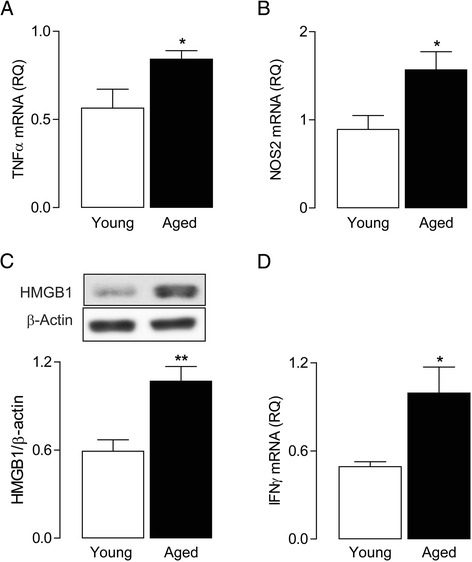
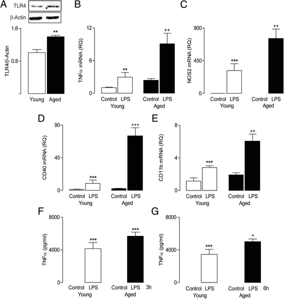
Background: Lipopolysaccharide (LPS) and interferon-γ (IFNγ) increase expression of tumour necrosis factor-α (TNFα) that characterizes the M1 activation state of macrophages. Whereas it is accepted that the immune system undergoes changes with age, there is inconsistency in the literature with respect to the impact of age on the response of macrophages to inflammatory stimuli. Here, we investigate the effect of age on the responsiveness of bone marrow-derived macrophages (BMDMs) to LPS and IFNγ. The context for addressing this question is that macrophages, which infiltrate the brain of aged animals, will encounter the neuroinflammatory environment that has been described with age.

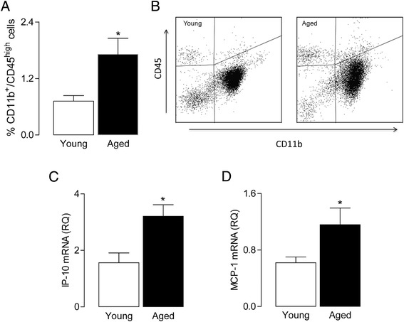
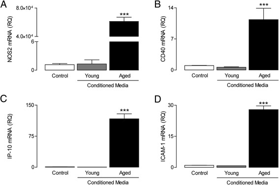
Methods: Brain tissue, prepared from young and aged rats, was assessed for expression of inflammatory markers by PCR and for evidence of infiltration of macrophages by flow cytometry. BMDMs were prepared from the long bones of young and aged rats, maintained in culture for 8 days and incubated in the presence or absence of LPS (100 ng/ml) or IFNγ (50 ng/ml). Cells were harvested and assessed for mRNA expression of markers of M1 activation including TNFα and NOS2, or for expression of IFNγR1 and TLR4 by western immunoblotting. To assess whether BMDMs induced glial activation, mixed glial cultures were incubated in the presence of conditioned media obtained from unstimulated BMDMs of young and aged rats and evaluated for expression of inflammatory markers.

Results: Markers associated with M1 activation were expressed to a greater extent in BMDMs from aged rats in response to LPS and IFNγ, compared with cells from young rats. The increased responsiveness was associated with increases in IFNγ receptor (IFNγR) and Toll-like receptor 4 (TLR4). The data show that conditioned media from BMDMs of aged rats increased the expression of pro-inflammatory mediators in glial cells. Significantly, there was an age-related increase in macrophage infiltration into the brain, and this was combined with increased expression of IFNγ and the Toll-like receptor 4 agonist, high-mobility group protein B1 (HMGB1).

Conclusion: Exposure of infiltrating macrophages to the inflammatory microenvironment that develops in the brain with age is likely to contribute to a damaging cascade that negatively impacts neuronal function.


Figures

Figure 1
Figure 1
Enhanced pro-inflammatory profile in the aged brain. Hippocampal expression of TNFα (A), NOS2 (B) and IFNγ (D) mRNA was significantly greater in aged rats (* P < 0.05; Student’s t-test; n = 5 to 15). Age was also associated with an increase in HMGB1 (C) protein expression in hippocampal tissue (** P < 0.01; Student’s t-test; n = 5 to 7). For mRNA data, values are presented as means (±SEM) and expressed as a ratio to β-actin mRNA. HMGB1, high-mobility group protein B1; IFNγ, interferon-γ; NOS2, nitric oxide synthase; RQ, relative quantities; TNFα, tumour necrosis factor-α.
Figure 2
Figure 2
Age is associated with increased macrophage infiltration and chemokine expression. There was a significant increase in the percentage of macrophages (A) in the brains of older rats (* P < 0.05; Student’s t-test; n = 4); values are presented as means (±SEM). (B) A representative plot of the flow cytometry data. There was an age-related increase in IP-10 (C) and MCP-1 (D) mRNA expression in hippocampal tissue (* P < 0.05; Student’s t-test; n = 6 to 15). For mRNA data, values are presented as means (±SEM) and expressed as a ratio to β-actin mRNA. IP-10, interferon gamma-induced protein 10; MCP-1, monocytes chemotactic protein 1; RQ, relative quantities.
Figure 3
Figure 3
Age is associated with increased TLR4 expression and enhanced sensitivity to LPS. TLR4 (A) protein expression (assessed by western immunoblot) in BMDMs was found to be increased in an age-related manner (** P < 0.01; Student’s t-test; n = 3). LPS (100 ng/ml; 24 h) stimulation significantly increased TNFα (B), NOS2 (C), CD40 (D) and CD11b (E) mRNA in BMDMs from young and aged rats (*** P < 0.001; ** P < 0.01; ANOVA); the LPS-induced increase in mRNA expression of these markers was significantly greater in BMDMs cultured from aged, compared with young, rats (++ P < 0.01; +++ P < 0.001; ANOVA). LPS stimulation (100 ng/ml; 3 and 6 h) significantly increased supernatant concentration of TNFα (measured by ELISA) in BMDMs from young and aged rats (F, G). While significant TNFα release was identified in cells from young and aged animals (F, G; *** P < 0.001, ANOVA), this LPS effect was significantly greater in BMDMs from aged rats at 6 h (G; + P < 0.05, ANOVA). Values are presented as means (±SEM; n = 3) expressed as a ratio to β-actin mRNA or as pg/ml. LPS, lipopolysaccharide; NOS2, nitric oxide synthase 2; CD40, cluster of differentiation 40; CD11b, cluster of differentiation 11b; TNFα, tumour necrosis factor-α; RQ, relative quantities.
Figure 4
Figure 4
IFNγ induces NOS2 and TNFα mRNA expression in macrophages from young and aged rats. IFNγR1 (A) protein expression (assessed by western immunoblot) was found to be greater in BMDMs prepared from aged compared with young rats (* P < 0.05, Student’s t-test; n = 3). IFNγ stimulation (50 ng/ml; 24 h) significantly increased TNFα (B), NOS2 (C), CD40 (D) and MHCII (E) mRNA in BMDMs from young and aged rats (*** P < 0.001; ANOVA). The IFNγ-induced increase in TNFα, CD40 and MHCII mRNA was significantly greater in BMDMs cultured from aged rats compared to cells derived from young rats (++ P < 0.01; +++ P < 0.001; ANOVA). (F, G) BMDMs were treated with IFNγ (50 ng/ml) for 3 and 6 h; following this, TNFα supernatant concentration was measured by ELISA. The IFNγ-induced TNFα release from BMDMs only reached significance in cells from aged rats following 3- and 6-h incubation (*P < 0.05, ANOVA; n = 3). Values are presented as means (±SEM; n = 3) expressed as a ratio to β-actin mRNA or as pg/ml. IFNγ, interferon-γ; IFNγR1, IFNγ receptor; NOS2, nitric oxide synthase; CD40, cluster of differentiation 40; MHCII, major histocampatability complex II; TNFα, tumour necrosis factor-α; RQ, relative quantities.
Figure 5
Figure 5
Conditioned media derived from aged BMDMs induces glial activation. Neonatal mixed glial cultures were incubated in the presence of conditioned media from unstimulated BMDMs derived from young and aged rats (24 h). Conditioned media from BMDMs derived from aged animals induced the expression of NOS2 (A), CD40 (B), IP-10 (C) and ICAM-1 mRNA ((D); *** P < 0.001; ANOVA), whereas exposure to conditioned media from BMDMs of young rats had no effect. Values are presented as means (±SEM; n = 3) expressed as a ratio to β-actin mRNA. NOS2, nitric oxide synthase; CD40, cluster of differentiation 40; IP-10, interferon gamma-induced protein 10; ICAM-1, intracellular adhesion molecule 1; RQ, relative quantities.

References

    1. Mosser DM, Zhang X. Activation of murine macrophages. Curr Protoc Immunol. 2008;Chapter 14:Unit 14.2. - PMC - PubMed
    1. Mosser DM. The many faces of macrophage activation. J Leukoc Biol. 2003;73:209–12. doi: 10.1189/jlb.0602325. - DOI - PubMed
    1. Stein M, Keshav S, Harris N, Gordon S. Interleukin 4 potently enhances murine macrophage mannose receptor activity: a marker of alternative immunologic macrophage activation. J Exp Med. 1992;176:287–92. doi: 10.1084/jem.176.1.287. - DOI - PMC - PubMed
    1. Gordon S, Martinez FO. Alternative activation of macrophages: mechanism and functions. Immunity. 2010;32:593–604. doi: 10.1016/j.immuni.2010.05.007. - DOI - PubMed
    1. Varin A, Gordon S. Alternative activation of macrophages: immune function and cellular biology. Immunobiology. 2009;214:630–41. doi: 10.1016/j.imbio.2008.11.009. - DOI - PubMed

Publication types