Review
. 2015 Feb 11;17(1):29.
doi: 10.1186/s13075-015-0542-4.

T-cell metabolism in autoimmune disease

Affiliations
Review

T-cell metabolism in autoimmune disease

Zhen Yang et al. Arthritis Res Ther. .

Abstract

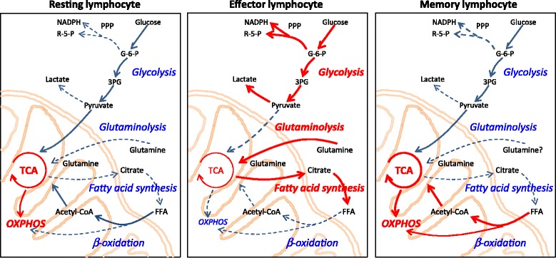
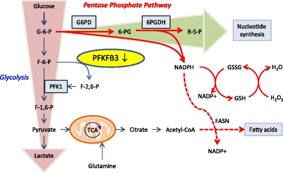
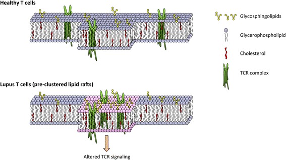
Cancer cells have long been known to fuel their pathogenic growth habits by sustaining a high glycolytic flux, first described almost 90 years ago as the so-called Warburg effect. Immune cells utilize a similar strategy to generate the energy carriers and metabolic intermediates they need to produce biomass and inflammatory mediators. Resting lymphocytes generate energy through oxidative phosphorylation and breakdown of fatty acids, and upon activation rapidly switch to aerobic glycolysis and low tricarboxylic acid flux. T cells in patients with rheumatoid arthritis (RA) and systemic lupus erythematosus (SLE) have a disease-specific metabolic signature that may explain, at least in part, why they are dysfunctional. RA T cells are characterized by low adenosine triphosphate and lactate levels and increased availability of the cellular reductant NADPH. This anti-Warburg effect results from insufficient activity of the glycolytic enzyme phosphofructokinase and differentiates the metabolic status in RA T cells from those in cancer cells. Excess production of reactive oxygen species and a defect in lipid metabolism characterizes metabolic conditions in SLE T cells. Owing to increased production of the glycosphingolipids lactosylceramide, globotriaosylceramide and monosialotetrahexosylganglioside, SLE T cells change membrane raft formation and fail to phosphorylate pERK, yet hyperproliferate. Borrowing from cancer metabolomics, the metabolic modifications occurring in autoimmune disease are probably heterogeneous and context dependent. Variations of glucose, amino acid and lipid metabolism in different disease states may provide opportunities to develop biomarkers and exploit metabolic pathways as therapeutic targets.


Figures

Figure 1
Figure 1
Metabolic pathways match T cells’ functional demands. Schematic diagrams of metabolic pathways employed by T cells at different stages of activation and differentiation. Dominant pathways are indicated as red cascades. Blue arrows show pathways that are used at a steady level, and dashed arrows indicate pathways that might be utilized but are insufficiently investigated. (Left) Resting lymphocytes generate energy from glucose, fatty acids and amino acids. Most ATP is produced in mitochondria by fermentation of acetyl-coenzyme A (CoA) in the tricarboxylic acid (TCA) cycle and oxidative phosphorylation (OXPHOS). (Middle) Effector lymphocytes (activated lymphocytes) swiftly and massively upregulate glycolysis and glutaminolysis, while keeping the TCA cycle low. These cells switch lipid metabolism from beta-oxidation towards fatty acid synthesis (lipogenesis). (Right) Memory lymphocytes mainly use beta-oxidation to support their energy needs. 3PG, 3-phosphoglycerate; FFA, free fatty acid; G-6-P, glucose-6-phosphate; NADPH, nicotinamide adenine dinucleotide phosphate; PPP, pentose phosphate pathway; R-5-P, ribose 5-phosphate.
Figure 2
Figure 2
Metabolic reprogramming in rheumatoid arthritis T cells. In contrast to healthy CD4 T cells, rheumatoid arthritis T cells fail to upregulate glycolytic activity due to the insufficient induction of 6-phosphofructo-2-kinase/fructose-2,6-bisphosphatase 3 (PFKFB3), a key regulatory enzyme in the glycolytic pathway. Deficient activity of PFKFB3 shunts glucose towards the pentose phosphate pathway and increases intracellular NADPH levels, hence unbalancing the cell’s redox status. 6PGDH, 6-phosphogluconate dehydrogenase; F-1,6-P, fructose-1,6-bisphosphatase; F-2,6-P, fructose-2,6-bisphosphatase; F-6-P, fructose-6-phosphate; FASN, fatty acid synthase; G-6-P, glucose-6-phosphate; G6PD, glucose-6-phosphate dehydrogenase; GSH, glutathione; GSSG, glutathione disulfide; NADPH, nicotinamide adenine dinucleotide phosphate; PFK, 6-phosphofructo-2-kinase; R-5-P, ribose 5-phosphate; TCA, tricarboxylic acid.
Figure 3
Figure 3
Altered membrane lipids in lupus T cells. The amount of glycerophospholipid, glycosphingolipids and cholesterol is tightly regulated and critical for T-cell receptor (TCR) signaling in healthy T cells. T cells from systemic lupus erythematosus patients exhibit excessive glycosphingolipid homeostasis, leading to aggregated lipid rafting and altered TCR signaling.

References

    1. Warburg O. On respiratory impairment in cancer cells. Science. 1956;124:269–70. - PubMed
    1. Hotamisligil GS. Inflammation and metabolic disorders. Nature. 2006;444:860–7. doi: 10.1038/nature05485. - DOI - PubMed
    1. Warburg O, Gawehn K, Geissler AW. Metabolism of leukocytes. Z Naturforsch B. 1958;13B:515–6. - PubMed
    1. Frauwirth KA, Riley JL, Harris MH, Parry RV, Rathmell JC, Plas DR, et al. The CD28 signaling pathway regulates glucose metabolism. Immunity. 2002;16:769–77. doi: 10.1016/S1074-7613(02)00323-0. - DOI - PubMed
    1. Sinclair LV, Rolf J, Emslie E, Shi YB, Taylor PM, Cantrell DA. Control of amino-acid transport by antigen receptors coordinates the metabolic reprogramming essential for T cell differentiation. Nat Immunol. 2013;14:500–8. doi: 10.1038/ni.2556. - DOI - PMC - PubMed

Publication types

MeSH terms