. 2015 Jun:53:149-59.
doi: 10.1016/j.biomaterials.2015.02.092. Epub 2015 Mar 13.

Acellular bi-layer silk fibroin scaffolds support functional tissue regeneration in a rat model of onlay esophagoplasty

Affiliations

Acellular bi-layer silk fibroin scaffolds support functional tissue regeneration in a rat model of onlay esophagoplasty

Khalid Algarrahi et al. Biomaterials. 2015 Jun.

Abstract

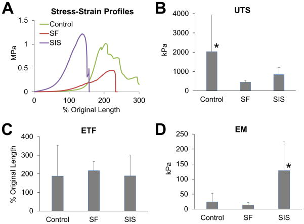
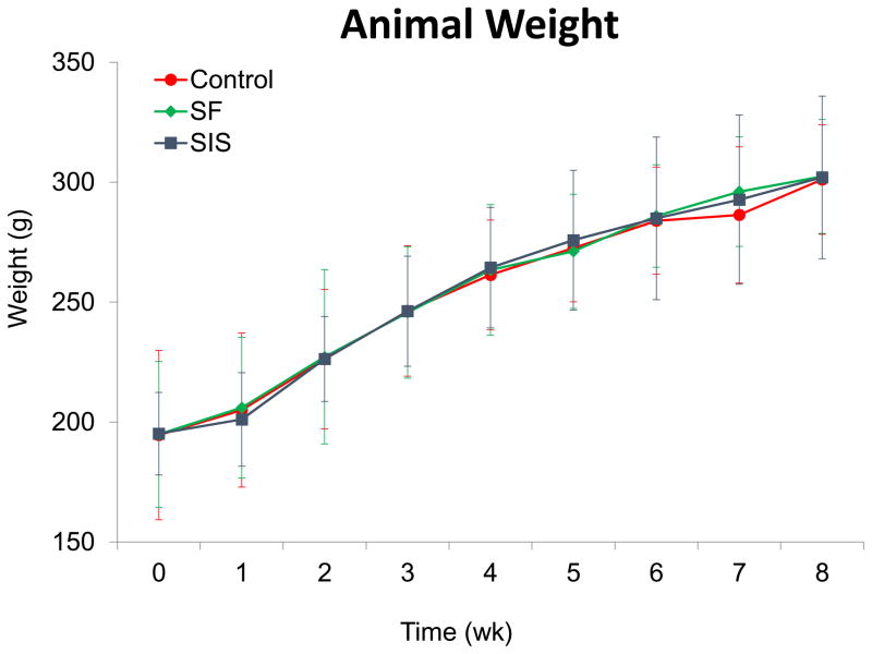
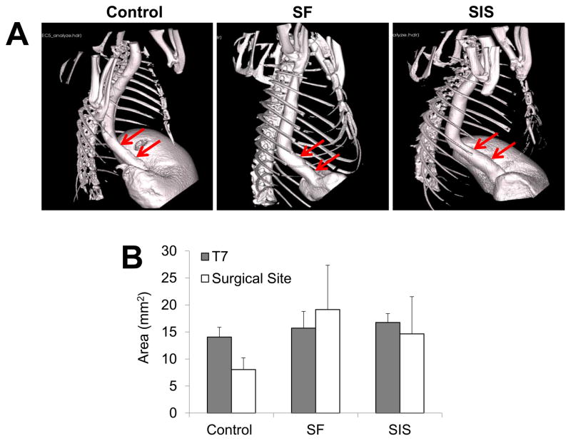
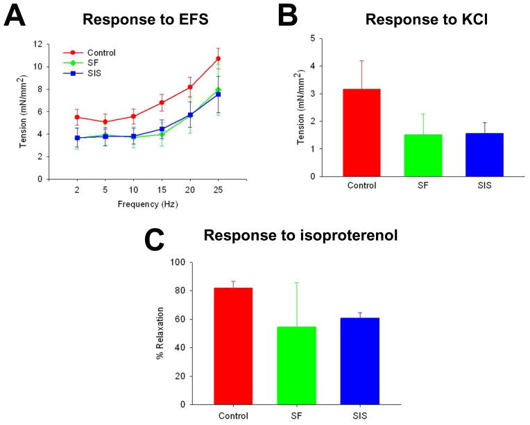
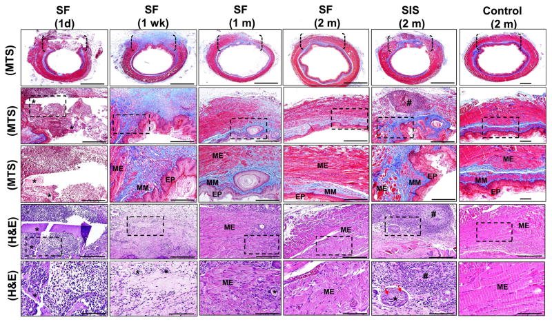
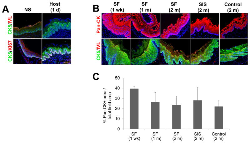
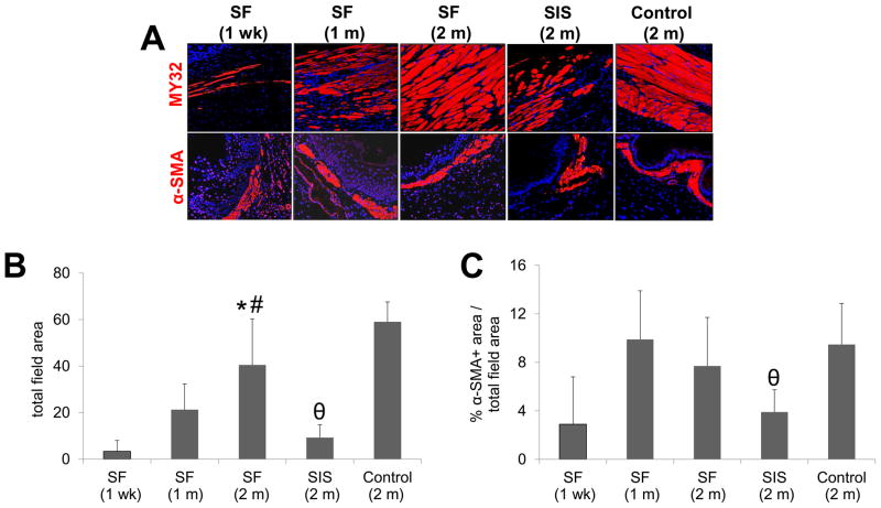
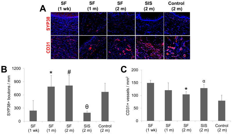
Surgical management of long-gap esophageal defects with autologous gastrointestinal tissues is frequently associated with adverse complications including organ dysmotility, dysphagia, and donor site morbidity. In order to develop alternative graft options, bi-layer silk fibroin (SF) scaffolds were investigated for their potential to support functional tissue regeneration in a rodent model of esophageal repair. Onlay esophagoplasty was performed with SF matrices (N = 40) in adult rats for up to 2 m of implantation. Parallel groups consisted of animals implanted with small intestinal submucosa (SIS) scaffolds (N = 22) or sham controls receiving esophagotomy alone (N = 20). Sham controls exhibited a 100% survival rate while rats implanted with SF and SIS scaffolds displayed respective survival rates of 93% and 91% prior to scheduled euthanasia. Animals in each experimental group were capable of solid food consumption following a 3 d post-op liquid diet and demonstrated similar degrees of weight gain throughout the study period. End-point μ-computed tomography at 2 m post-op revealed no evidence of contrast extravasation, fistulas, strictures, or diverticula in any of the implant groups. Ex vivo tissue bath studies demonstrated that reconstructed esophageal conduits supported by both SF and SIS scaffolds displayed contractile responses to carbachol, KCl and electrical field stimulation while isoproterenol produced tissue relaxation. Histological (Masson's trichrome and hematoxylin and eosin) and immunohistochemical (IHC) evaluations demonstrated both implant groups produced de novo formation of skeletal and smooth muscle bundles positive for contractile protein expression [fast myosin heavy chain (MY32) and α-smooth muscle actin (α-SMA)] within the graft site. However, SF matrices promoted a significant 4-fold increase in MY32+ skeletal muscle and a 2-fold gain in α-SMA+ smooth muscle in comparison to the SIS cohort as determined by histomorphometric analyses. A stratified squamous, keratinized epithelium expressing cytokeratin 5 and involucrin proteins was also present at 2 m post-op in all experimental groups. De novo innervation and vascularization were evident in all regenerated tissues indicated by the presence of synaptophysin (SYP38)+ boutons and vessels lined with CD31 expressing endothelial cells. In respect to SIS, the SF group supported a significant 4-fold increase in the density of SYP38+ boutons within the implant region. Evaluation of host tissue responses revealed that SIS matrices elicited chronic inflammatory reactions and severe fibrosis throughout the neotissues, in contrast to SF scaffolds. The results of this study demonstrate that bi-layer SF scaffolds represent promising biomaterials for onlay esophagoplasty, capable of producing superior regenerative outcomes in comparison to conventional SIS scaffolds.

Keywords: Epithelium; Muscle; Scaffold; Silk; Wound healing.


Figures

Figure 1
Figure 1. Rat onlay esophagoplasty model
Photomicrographs of various surgical stages of scaffold implantation and gross morphology of regenerated tissues. [A] Esophagotomy and exposure of the esophagus lumen. [B] Anastomosis of bi-layer silk fibroin (SF) graft into the esophagus defect. [C, D] Regenerated tissues present within the original implantation sites supported by SF scaffolds [C] and small intestinal submucosa (SIS) matrices [D] at 2 m post-op. Arrows denote original marking sutures. Scale bars = 7 mm.
Figure 2
Figure 2. Tensile properties of regenerated esophageal tissues supported by implant and sham control groups
[A] Representative stress-strain profiles of experimental groups at 2 m post-op. [BD] Evaluation of ultimate tensile strength (UTS), elongation to failure (ETF), and elastic modulus (EM) in cohorts defined in [A]. Means ± standard deviation per data point. (*) = p<0.05 in comparison to all other groups.
Figure 3
Figure 3. Body weight evaluations in matrix-grafted animals and sham controls both pre-operatively and over the course of the study period
All experimental subjects were fed a liquid diet for 3 d post-op and subsequently maintained on solid food thereafter. Means ± standard deviation per data point.
Figure 4
Figure 4. μCT analysis of esophageal continuity in implant and sham control groups
[A] Representative 3-D images of esophagi in experimental groups at 2 m post-op following contrast gavage. Arrows denote boundaries of original graft anastomosis or esophagotomy in controls. [B] Quantification of luminal esophageal cross-sectional areas from the central region of the original scaffold anastomosis or esophagotomy in controls (surgical sites) as well as a nonsurgical reference point adjacent to the 7th thoracic vertebra (T7). Means ± standard deviation per data point.
Figure 5
Figure 5. Ex vivo tissue contractility and relaxation responses in reconstructed esophageal conduits isolated from matrix and sham control groups
[A] Frequency response curves to electrical field stimulation (EFS) in circular esophageal rings containing original graft sites or esophagotomy region following 2 m post-op. [B] Contractile responses to KCl (80 mM) in specimens described in [A]. [C] Relaxation responses in samples detailed in [A] in response to isoproterenol (10 μM) following pre-contraction with carbachol (1 μM). Means ± standard deviation per data point.
Figure 6
Figure 6. Histological evaluations of tissue regeneration and host responses in sham controls and implant groups
[1st row] Photomicrographs of MTS-stained gross esophageal cross-sections containing region of tissue repair. Brackets denote sites of scaffold anastomosis or control esophagotomy. Scale bars = 1.25 mm. [2nd row] Magnification of global tissue regeneration area (RA) bracketed in 1st column. Scale bars = 750 μm. [3rd row] Magnified boxed area in 2nd column. Scale bars = 250 μm. [4th row] Photomicrographs of H&E-stained sections from bracketed area described in 1st column. Scale bars = 250 μm. [5th row] Magnified boxed area in 4th row. Scale bars = 80 μm. EP = epithelium; MM = muscularis mucosa; ME = muscularis externa. (*) = scaffold remnants. (#) = aggregate of mononuclear cells indicative of chronic inflammation. Red arrows denote multi-nuclear giant cells encapsulating residual scaffold remnants.
Figure 7
Figure 7. Immunohistochemical and histomorphometric assessments of epithelial regeneration in controls and scaffold groups
[A] Photomicrographs of involucrin (IVL), cytokeratin 5 (CK5), and Ki67 protein expression in epithelia present in nonsurgical (NS) controls and in host tissue adjacent to the site of SF scaffold anastomosis. [B] Photomicrographs of pan-CK, IVL, and CK5 protein expression in de novo epithelia present within the original graft sites or control esophagotomy area. For all panels, respective marker expression is displayed in red (Cy3) or green (FITC) labeling. Blue denotes DAPI nuclear counterstain. Scale bars in all panels = 200 μm. [C] Histomorphometric analysis of the extent of pan-CK+ epithelia present at site of tissue repair in scaffold groups or sham-operated controls.
Figure 8
Figure 8. Immunohistochemical and histomorphometric evaluations of skeletal and smooth muscle formation in sham controls and scaffold groups
[A] Photomicrographs of fast myosin skeletal heavy chain (MY32) and α-smooth muscle actin (α-SMA) protein expression in de novo muscularis externa and muscularis muscosa, respectively, within the original graft site or control esophagotomy area. For all panels, respective marker expression is displayed in red (Cy3) labeling. Blue denotes DAPI nuclear counterstain. Scale bars in all panels = 200 μm. [B, C] Histomorphometric analysis of the extent of MY32+ skeletal muscle [B] and α-SMA+ smooth muscle [C] present at sites of tissue repair in matrix groups or sham controls. (*) = p<0.05 in comparison to both 1 wk and 1 m SF groups. (#) = p<0.05 in comparison to SIS. (θ) = p<0.05 in comparison to sham control.
Figure 9
Figure 9. Immunohistochemical and histomorphometric analyses of de novo innervation and vascularization in experimental groups
[A] Photomicrographs of synaptophysin (SYP38) and CD31 protein expression present within original graft sites or control esophagotomy area. For all panels, respective marker expression is displayed in red (Cy3) labeling. Blue denotes DAPI nuclear counterstain. Arrows denote SYP38+ boutons. Scale bars in all panels = 200 μm. [B, C] Histomorphometric analysis of density of SYP38+ boutons [B] and CD31+ vessels [C] observed at sites of tissue regeneration in implant groups or sham controls. (*) = p<0.05 in comparison to 1 wk SF group. (#) = p<0.05 in comparison to SIS. (θ) = p<0.05 in comparison to sham control. (α) = p<0.05 in comparison to all other 2 m groups.

References

    1. Shen KR, Austen WG, Jr, Mathisen DJ. Use of a prefabricated pectoralis major muscle flap and pedicled jejunal interposition graft for salvage esophageal reconstruction after failed gastric pull-up and colon interposition. J Thorac Cardiovasc Surg. 2008;135:1186–7. - PubMed
    1. Burgos L, Barrena S, Andrés AM, Martínez L, Hernández F, Olivares P, et al. Colonic interposition for esophageal replacement in children remains a good choice: 33-year median follow-up of 65 patients. J Pediatr Surg. 2010;45:341–5. - PubMed
    1. Leonard GD, McCaffrey JA, Maher M. Optimal therapy for oesophageal cancer. Cancer Treat Rev. 2003;29:275–82. - PubMed
    1. Greene CL, DeMeester SR, Worrell SG, Oh DS, Hagen JA, DeMeester TR. Alimentary satisfaction, gastrointestinal symptoms, and quality of life 10 or more years after esophagectomy with gastric pull-up. J Thorac Cardiovasc Surg. 2014;147:909–14. - PubMed
    1. Romeo C, Bonanno N, Baldari S, Centorrino A, Scalfari G, Antonuccio P, et al. Gastric motility disorders in patients operated on for esophageal atresia and tracheoesophageal fistula: long term evaluation. J Pediatr Surg. 2000;35:740–4. - PubMed

Publication types