. 2015:53:437-52.
doi: 10.1016/j.biomaterials.2015.02.083. Epub 2015 Mar 19.

The interaction of carbon nanotubes with an in vitro blood-brain barrier model and mouse brain in vivo

Affiliations

The interaction of carbon nanotubes with an in vitro blood-brain barrier model and mouse brain in vivo

Houmam Kafa et al. Biomaterials. 2015.

Abstract

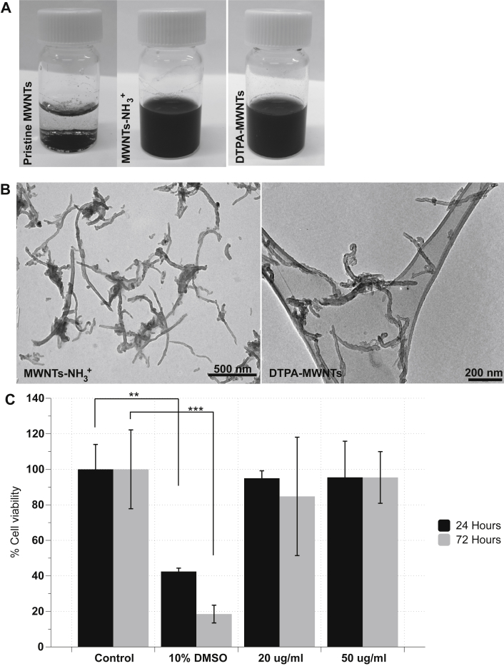
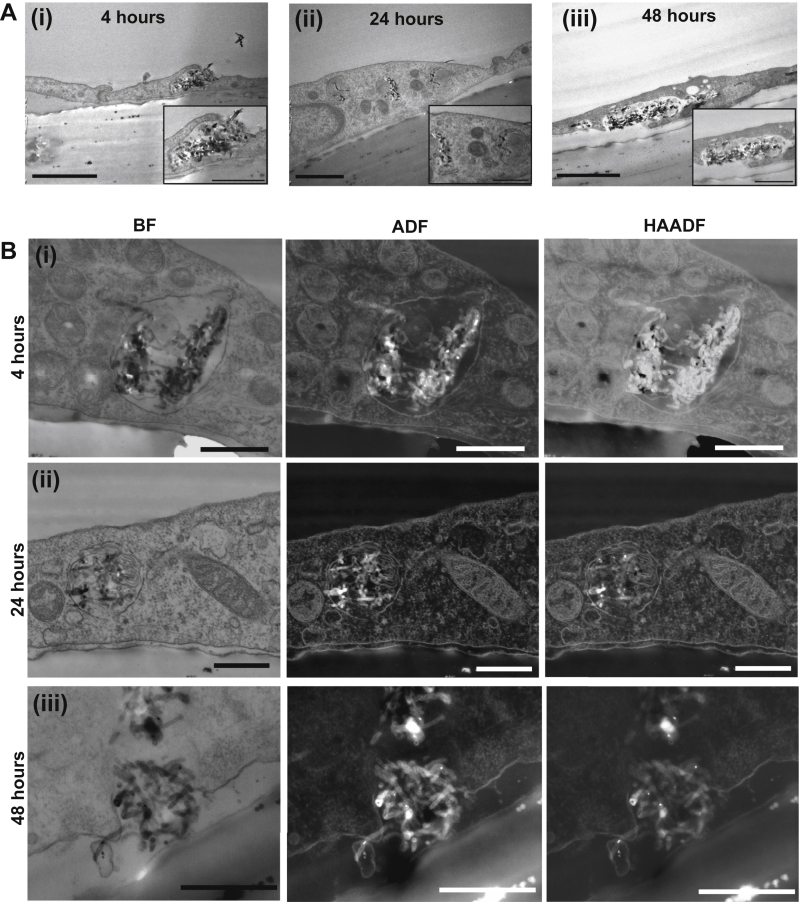
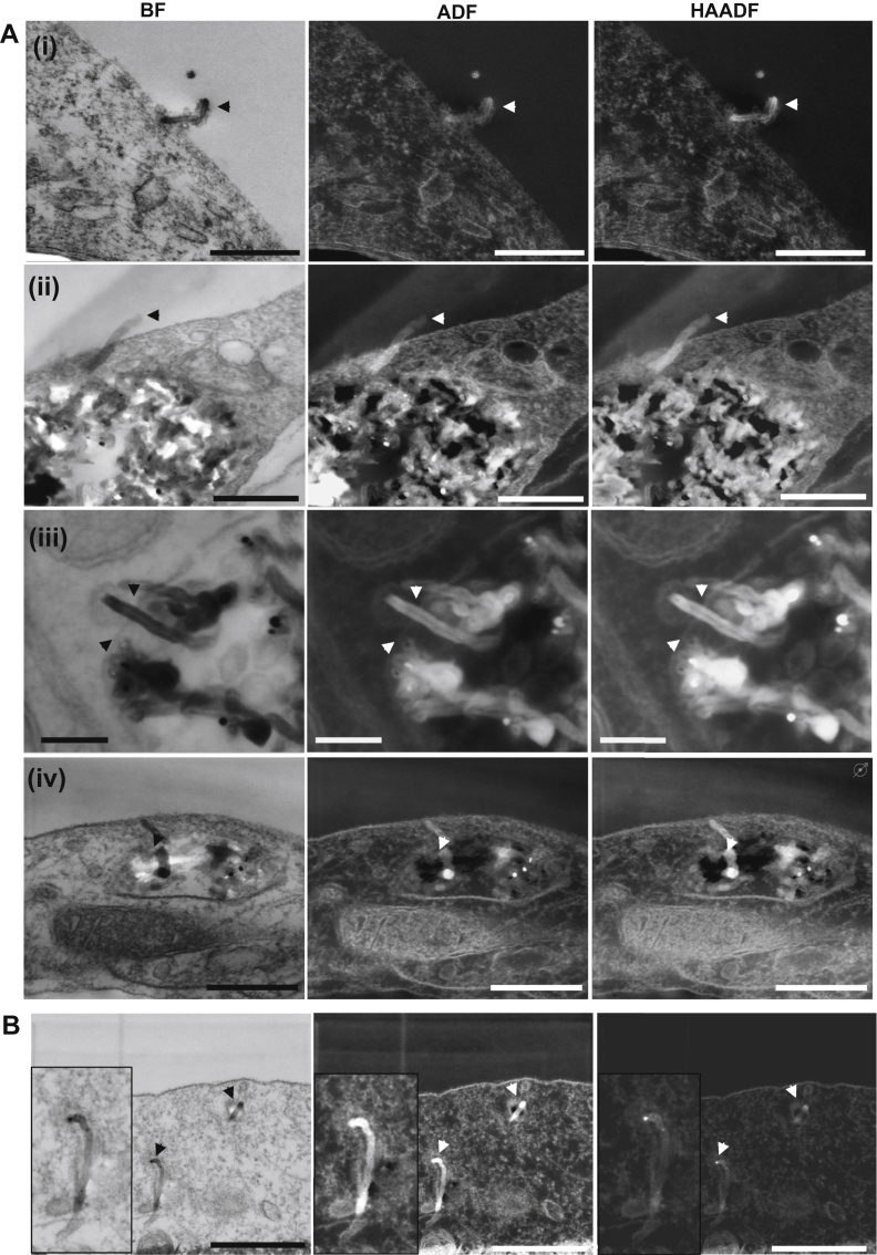
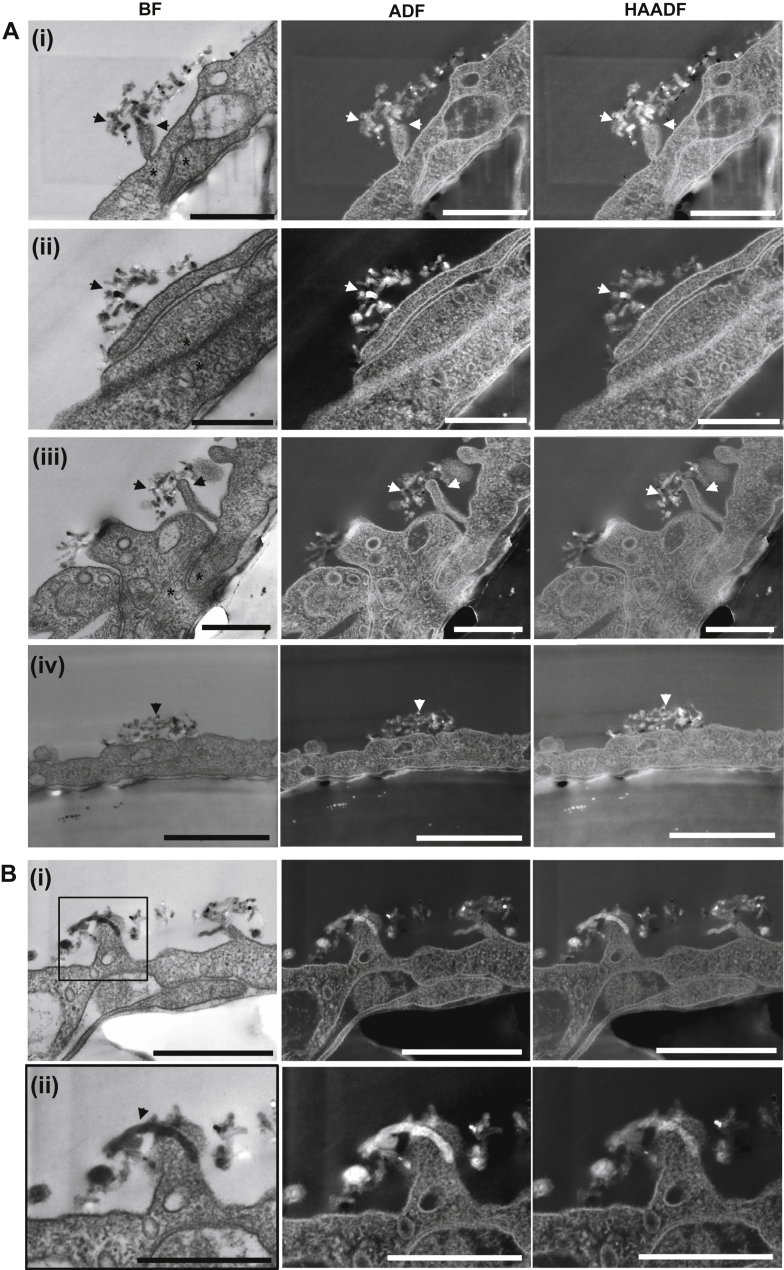
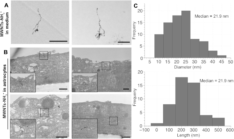
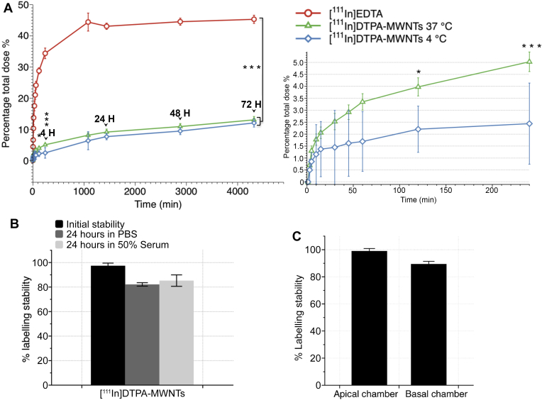
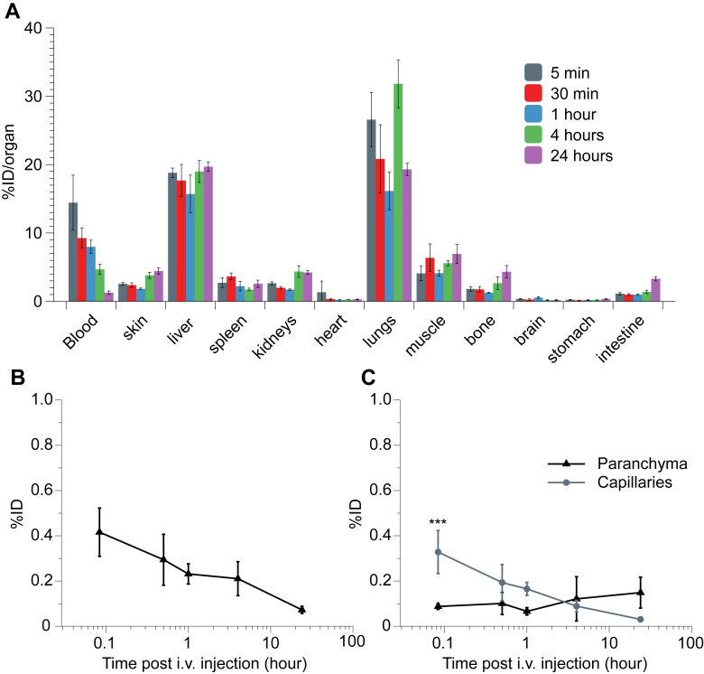
Carbon nanotubes (CNTs) are a novel nanocarriers with interesting physical and chemical properties. Here we investigate the ability of amino-functionalized multi-walled carbon nanotubes (MWNTs-NH3(+)) to cross the Blood-Brain Barrier (BBB) in vitro using a co-culture BBB model comprising primary porcine brain endothelial cells (PBEC) and primary rat astrocytes, and in vivo following a systemic administration of radiolabelled f-MWNTs. Transmission Electron microscopy (TEM) confirmed that MWNTs-NH3(+) crossed the PBEC monolayer via energy-dependent transcytosis. MWNTs-NH3(+) were observed within endocytic vesicles and multi-vesicular bodies after 4 and 24 h. A complete crossing of the in vitro BBB model was observed after 48 h, which was further confirmed by the presence of MWNTs-NH3(+) within the astrocytes. MWNT-NH3(+) that crossed the PBEC layer was quantitatively assessed using radioactive tracers. A maximum transport of 13.0 ± 1.1% after 72 h was achieved using the co-culture model. f-MWNT exhibited significant brain uptake (1.1 ± 0.3% injected dose/g) at 5 min after intravenous injection in mice, after whole body perfusion with heparinized saline. Capillary depletion confirmed presence of f-MWNT in both brain capillaries and parenchyma fractions. These results could pave the way for use of CNTs as nanocarriers for delivery of drugs and biologics to the brain, after systemic administration.

Keywords: BBB model; PBEC; STEM; TEM; Transcytosis; Transwells.


Figures

Fig. 1
Fig. 1
The dispersibility of f-MWNTs constructs and the toxicity of MWNTs-NH3+on PBEC after 24 and 72 h of uptake. The water dispersibility of the functionalized MWNTs-NH3+ and DTPA-MWNTs in water as observed visually (A) and by TEM examination (B). (C) The modified LDH assay was carried out to evaluate cell viability after 24 and 72 h of incubation at 20 and 50 μg/ml. 10% DMSO (v/v) was used as positive control. MWNTs-NH3+ had minimal effect on cell viability after 24 and 72 h while the viability of the cells treated with 10% DMSO was significantly reduced compared to the control at both time points. MWNTs-NH3+ did not affect the viability of PBEC up to 50 μg/ml (one-way ANOVA test; *p < 0.05, **p < 0.01, ***p < 0.001, n = 3).
Fig. 2
Fig. 2
The transcytosis pattern of MWNTs-NH3+across the PBEC monolayer following the incubation of MWNTs-NH3+with an in vitro BBB model. MWNTs-NH3+ (20 μg/ml) were added to the apical chamber and incubated with the PBEC for (i) 4, (ii) 24 and (iii) 48 h. (A) Bright field TEM images of polyester filters showing the initial interaction of MWNTs-NH3+ with the PBEC monolayer after 4 h (i). After 24 h (ii), the MWNTs-NH3+ clusters appeared within endocytic vesicles or multi-vesicular bodies. The MWNTs-NH3+-containing vesicles showed evidence of fusion with the abluminal plasma membrane, and were partly open towards the basal chamber After 48 h (iii). (B) Low voltage STEM images of the polyester filters confirming the uptake and transcytosis of MWNTs-NH3+ across the PBEC monolayer. MWNTs-NH3+ appeared within endocytic vesicles after (i) 4 and (ii) 24 h. The MWNTs-NH3+-containing vesicles were then imaged partly open towards the basal chamber allowing the release of MWNTs-NH3+ after 48 h (iii). The three detection modes: bright field (BF), annular dark field (ADF) and high angular annular dark field (HAADF) helped identifying the electron dense structures of MWNTs-NH3+ within the cell body, and the osmium-rich lipid membranes surrounding the nanostructures. Scale bars: (A) 1 μm and (inset) 500 nm. (B) First and second row 500 nm and last row 400 nm.
Fig. 3
Fig. 3
Translocation of “individual” MWNTs-NH3+across the PBEC membrane. Individual MWNTs-NH3+ shows an apparent ability to pierce through the plasma membrane. (A) Bright field STEM images showing a single MWNTs-NH3+ (black and white arrow heads) crossing the plasma membrane of endothelial cells (i, ii). The ADF and HAADF images clearly demonstrate part of the nanotube on the other side of the plasma membrane. The ability of MWNTs-NH3+ to permeate through plasma membranes was not only demonstrated at the plasma membrane level, but also seen as a transport method within the cell (iii, iv). (B) Electron micrographs showing a single nanotube that does not appear to be associated with other sub-cellular compartments. Close examination reveals the presence of a thin membrane around the single nanotube. Scale bars (A) from top to bottom, 400 nm, 300 nm and 100 nm, 300 nm. (B) 500 nm.
Fig. 4
Fig. 4
Inhibition of MWNTs-NH3+uptake into endothelial cells following the incubation of MWNTs-NH3+at 4 °C. (A) The transport experiment was carried out at 4 °C for 4 h to evaluate the energy dependency of MWNTs-NH3+ uptake into the endothelial cells. Low voltage STEM images show the accumulation of MWNTs-NH3+ outside the endothelial cells after 4 h of incubation, but no evidence of vesicular uptake was observed. The images confirmed that transcytosis of MWNTs-NH3+ was governed by an active mechanism. The MWNTs-NH3+ were mostly observed around endothelial extensions (arrow heads) in the vicinity of a tight-junctional zone (asterisks) (iiii). (B) Electron micrographs showing a single MWNTs-NH3+ interacting with the endothelial extension at 4 °C (i, ii). Part of the MWNTs-NH3+ appeared on the other side of the plasma membrane. Scale bars (A) from top to bottom, 500 nm, 400 nm and 500 nm, 1 μm. (B) From top to bottom 500 nm and 300 nm.
Fig. 5
Fig. 5
Uptake of MWNTs-NH3+into primary astrocytes following PBEC transcytosis. Electron micrographs showing the fate of MWNTs-NH3+ following the crossing of the PBEC monolayer. MWNTs-NH3+ were tracked into the basal chamber after 24 h of incubation with PBEC. (A) The structure of MWNTs-NH3+ in the medium of the basal chamber following crossing the PBEC/filter. (B) Electron micrographs showing the uptake of MWNTs-NH3+ into the primary astrocytes. Individual MWNTs-NH3+ were observed within the astrocytes with no evidence of vesicular uptake. (C) Length and diameter distribution histogram (n = 100) of the studied MWNTs-NH3+. The median length and diameter of the MWNTs-NH3+ was 233.9 nm and 21.9 nm, respectively. Scale bars = 500 nm.
Fig. 6
Fig. 6
The percentage of [111In]DTPA-MWNTs transported across the PBEC monolayer over 72 h. [111In]DTPA-MWNTs (20 μg/ml) were added to the apical chamber and incubated with PBEC at 37 °C or 4 °C (for the initial 4 h) followed by 37 °C incubation up to 72 h. The radioactivity in the basal chamber was measured in 0.5 ml aliquots at different time points (A). [111In]EDTA was used as a control. [111In]DTPA-MWNTs incubated at 37 °C crossed the PBEC monolayer and were detected in the basal chamber reaching a maximum of 13.0 ± 1.1% after 72 h. The incubation at 4 °C significantly decreased the uptake of [111In]DTPA-MWNTs at 2 h (p < 0.05) and 4 h (p < 0.001) which then increased to the same level as those at 37 °C, reaching a maximum of 12.0 ± 1.5%. The uptake of [111In]EDTA showed significantly higher permeation at all time points studied compared to [111In]DTPA-MWNTs, reaching a maximum of 45.3 ± 1.3% after 72 h (p < 0.0001). Inset to the right shows the difference in [111In]DTPA-MWNTs uptake during the initial 4 h of incubation. (One way ANOVA test; *p < 0.05, ***p < 0.001, ****p < 0.0001, n = 3). (B) the stability of the radioactive tag on the [111In]DTPA-MWNTs as measured by TLC using 0.1 M ammonium acetate buffer containing 50 mM EDTA. Radio-labelled [111In]DTPA-MWNTs were incubated with PBS or 50% serum at 37 °C for 24 h prior chromatographic analysis. (C) The radiolabelling stability of [111In]DTPA-MWNTs during the time course of the transport experiments measured in the apical and basal chamber of the co-culture model, respectively.
Fig. 7
Fig. 7
Organ biodistribution and brain uptake of [111In]DTPA-MWNTs into mice following i.v. administration. C57/Bl6 mice were injected with [111In]DTPA-MWNTs (50 μg, 0.5 MBq) via the tail vein. At each time point (5 min, 30 min, 1 h, 4 h and 24 h) whole body perfusion with heparinized saline was performed and major organs were harvested for quantitative measurements of radioactivity by γ-scintigraphy. (A) The accumulation of [111In]DTPA-MWNTs in the major organs after each time point. (B) Brain uptake of [111In]DTPA-MWNTs over time showing the highest brain accumulation after 5 min. (C) The capillary depletion profile showing the sustained accumulation of [111In]DTPA-MWNTs in brain parenchyma over time compared to a gradual reduction in capillary fraction. The data is presented as % injected dose per organ. Results are expressed as mean ± SD, n = 3 (***p < 0.001).

References

    1. Saito N., Usui Y., Aoki K., Narita N., Shimizu M., Hara K. Carbon nanotubes: biomaterial applications. Chem Soc Rev. 2009;38:1897–1903. - PubMed
    1. Bianco A., Kostarelos K., Prato M. Applications of carbon nanotubes in drug delivery. Curr Opin Chem Biol. 2005;9:674–679. - PubMed
    1. Cellot G., Cilia E., Cipollone S., Rancic V., Sucapane A., Giordani S. Carbon nanotubes might improve neuronal performance by favouring electrical shortcuts. Nat Nanotechnol. 2009;4:126–133. - PubMed
    1. Lee H.J., Park J., Yoon O.J., Kim H.W., Lee do Y., Kim do H. Amine-modified single-walled carbon nanotubes protect neurons from injury in a rat stroke model. Nat Nanotechnol. 2011;6:121–125. - PMC - PubMed
    1. Al-Jamal K.T., Gherardini L., Bardi G., Nunes A., Guo C., Bussy C. Functional motor recovery from brain ischemic insult by carbon nanotube-mediated siRNA silencing. Proc Natl Acad Sci U. S. A. 2011;108:10952–10957. - PMC - PubMed

Publication types