Review
. 2015 Sep 20;23(9):734-46.
doi: 10.1089/ars.2015.6247. Epub 2015 Jun 1.

The Redox Code

Affiliations
Review

The Redox Code

Dean P Jones et al. Antioxid Redox Signal. .

Abstract

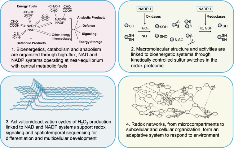
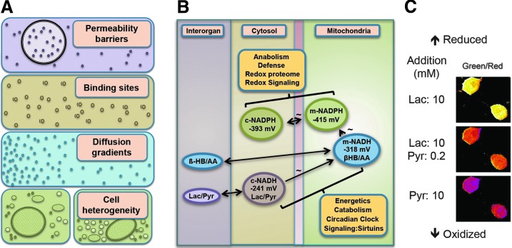
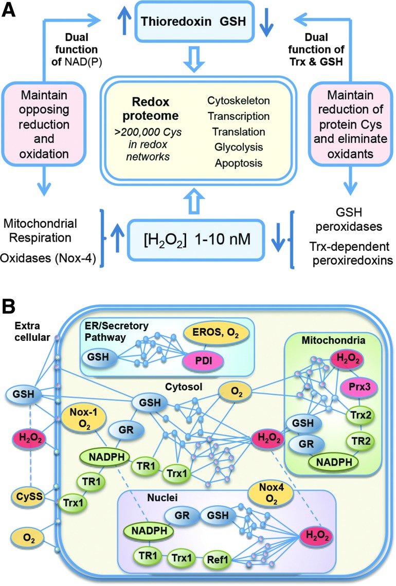
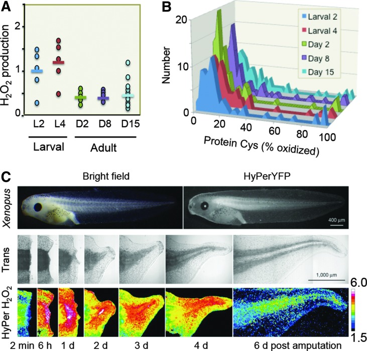
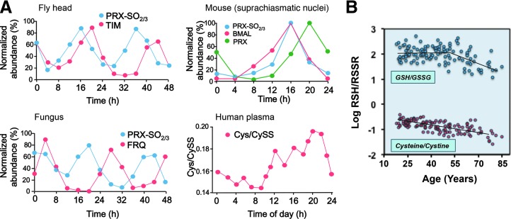
Significance: The redox code is a set of principles that defines the positioning of the nicotinamide adenine dinucleotide (NAD, NADP) and thiol/disulfide and other redox systems as well as the thiol redox proteome in space and time in biological systems. The code is richly elaborated in an oxygen-dependent life, where activation/deactivation cycles involving O₂ and H₂O₂ contribute to spatiotemporal organization for differentiation, development, and adaptation to the environment. Disruption of this organizational structure during oxidative stress represents a fundamental mechanism in system failure and disease.

Recent advances: Methodology in assessing components of the redox code under physiological conditions has progressed, permitting insight into spatiotemporal organization and allowing for identification of redox partners in redox proteomics and redox metabolomics.

Critical issues: Complexity of redox networks and redox regulation is being revealed step by step, yet much still needs to be learned.

Future directions: Detailed knowledge of the molecular patterns generated from the principles of the redox code under defined physiological or pathological conditions in cells and organs will contribute to understanding the redox component in health and disease. Ultimately, there will be a scientific basis to a modern redox medicine.


Figures

<b>FIG. 1.</b>
FIG. 1.
The redox code. The four principles of the redox code by which biological systems are organized (see text).
<b>FIG. 2.</b>
FIG. 2.
Metabolism is organized through high-flux NAD and NADP systems. (A) Multiple types of compartmentation affect free concentration of redox partners: microheterogeneity (permeability barriers, binding sites, diffusion gradients) and macroheterogeneity. (B) The NAD and NADP systems are maintained at steady states near thermodynamic equilibrium in mitochondria, cytosol, and other subcellular redox spaces to control energetics, catabolism, and anabolism. The cytoplasmic and mitochondrial NAD systems are respectively equilibrated throughout the body through lactate (Lac)/pyruvate (Pyr) and β-hydroxybutyrate (HB)/acetoacetate (AA). Mitochondrial NADH and NADPH systems are connected through energy-linked transhydrogenase. The midpoint potentials (two-electron half-cell reduction potentials) are given in mV. (C) Cytosolic NADH/NAD+ redox state imaged in cells with a genetically encoded fluorescent biosensor, Peredox, calibrated using exogenous lactate (Lac) and pyruvate (Pyr). (A, B) Modified from Sies (100, 101); (C) from Hung et al. (46) with permission of the publisher.
<b>FIG. 3.</b>
FIG. 3.
The redox proteome is organized through kinetically controlled sulfur switches linked to NAD and NADP systems. (A) Redox states of Cys in functional networks are maintained by opposing oxidation and reduction reactions. NADPH is the principal reductant used to maintain the redox states of the Trx and GSH systems. These systems maintain the redox proteome, with GSH functioning through glutaredoxins (not shown). Trx and GSH also regulate H2O2 concentrations through Prx and GSH peroxidases, respectively. H2O2 is generated metabolically by mitochondria and about 25 different oxidases. Nox-4 is included because of constitutive activity with NADPH and O2; different Nox systems support a range of signaling activities, while other H2O2-producing enzymes support a range of biosynthetic and detoxification functions. (B) Protein Cys in subcellular compartments is regulated by oxidation (red) and reduction (blue/green) to maintain organization structure through control of distribution, interaction, and activity. Cys, cysteine; CySS, cystine; EROS, endoplasmic reticulum oxidase system; GR, glutathione disulfide reductase; GSH, glutathione; Nox, NADPH oxidase; PDI, protein disulfide isomerase; Prx, peroxiredoxin; Ref-1, redox factor-1 (AP endonuclease-1); TR, thioredoxin reductase; Trx, thioredoxin.
<b>FIG. 4.</b>
FIG. 4.
Activation/deactivation cycles of H2O2 production support redox signaling and spatiotemporal organization of complex multicellular systems. (A) Redox imaging using HyPer reveals temporal sequence of H2O2 production in Caenorhabditis elegans development. Endogenous hydroperoxide levels during the life span of C. elegans were measured with HyPer ratios in the body wall muscle cells at different stages during their life span. Each symbol represents the HyPer ratio of an individual animal; the bar illustrates the average HyPer ratio per day. Experiments were performed a minimum of three times, and a representative graph shown here is redrawn from the original. One-way ANOVA, followed by the Tukey multiple comparison test, was performed on the log-transformed HyPer ratio and showed that the larval levels were different from the adult levels (p<0.05). Results were independently confirmed by Amplex UltraRed measurement of H2O2, which were also significantly different. (B) Protein Cys oxidation occurs in association with H2O2 production in C. elegans development. A complete list of proteins and standard deviations are given as an online supplement (http://dx.doi.org/10.1016/j.molcel.2012.06.016) to the original publication (58). (C) Redox imaging reveals the spatiotemporal sequence of H2O2 production in Xenopus laevis wound healing. Sequence of H2O2 production in tadpole tail regeneration is shown for days postamputation in the X. laevis line expressing HyPerYFP. The numbers give the excitation ratio of HyPerYFP490nm/HyPerYFP402nm, and the color scale reflects this ratio. (A, B) Modified from Knoefler et al. (58) and (C) from Love et al. (64), with permission from the publisher.
<b>FIG. 5.</b>
FIG. 5.
Redox network structure supports adaptation to the environment. (A) Oxidation cycles for Prx are linked to clock protein expression. The oxidation cycle for peroxiredoxin-6 (Prx-SO2/3) is shown for fly (Drosophila melanogaster) with clock protein timeless (TIM), fungus (Neurospora crassa) with clock protein (FRQ), and mouse (Mus musculus) Prx1 with circadian protein BMAL. Similar variation in Cys/CySS is shown for human plasma. Figures were redrawn and simplified from original publications; originals should be consulted for statistical measurements and additional details and validations. (B) Progressive oxidation with age, shown by human plasma Cys/CySS and GSH/GSSG, contributes to decline in adaptability. (A) Composite modified from Edgar et al. (24) and Blanco et al. (9); (B), modified from Jones et al. (49). BMAL, transcriptional activator, core component of circadian clock; GSSG, glutathione disulfide; Prx1, peroxiredoxin-1.

References

    1. Abate C, Patel L, Rauscher FJ, 3rd, and Curran T. Redox regulation of fos and jun DNA-binding activity in vitro. Science 249: 1157–1161, 1990 - PubMed
    1. Adimora NJ, Jones DP, and Kemp ML. A model of redox kinetics implicates the thiol proteome in cellular hydrogen peroxide responses. Antioxid Redox Signal 13: 731–743, 2010 - PMC - PubMed
    1. Babior BM, Kipnes RS, and Curnutte JT. Biological defense mechanisms. The production by leukocytes of superoxide, a potential bactericidal agent. J Clin Invest 52: 741–744, 1973 - PMC - PubMed
    1. Bass J. Circadian topology of metabolism. Nature 491: 348–356, 2012 - PubMed
    1. Beck MA, Nelson HK, Shi Q, Van Dael P, Shiffrin EJ, Blum S, Barclay D, and Levander OA. Selenium deficiency increases the pathology of an influenza infection. FASEB J 15: 1481–1483, 2001 - PubMed

Publication types