

Shared epitope-antagonistic ligands: a new therapeutic strategy in mice with erosive arthritis
- PMID: 25892196
- PMCID: PMC4784479
- DOI: 10.1002/art.39158
Shared epitope-antagonistic ligands: a new therapeutic strategy in mice with erosive arthritis
Abstract
Objective: The mechanisms underlying bone damage in rheumatoid arthritis (RA) are incompletely understood. We recently identified the shared epitope (SE), an HLA-DRB1-coded 5-amino acid sequence motif carried by the majority of RA patients as a signal transduction ligand that interacts with cell surface calreticulin and accelerates osteoclast (OC)-mediated bone damage in collagen-induced arthritis (CIA). Given the role of the SE/calreticulin pathway in arthritis-associated bone damage, we sought to determine the therapeutic targetability of calreticulin.
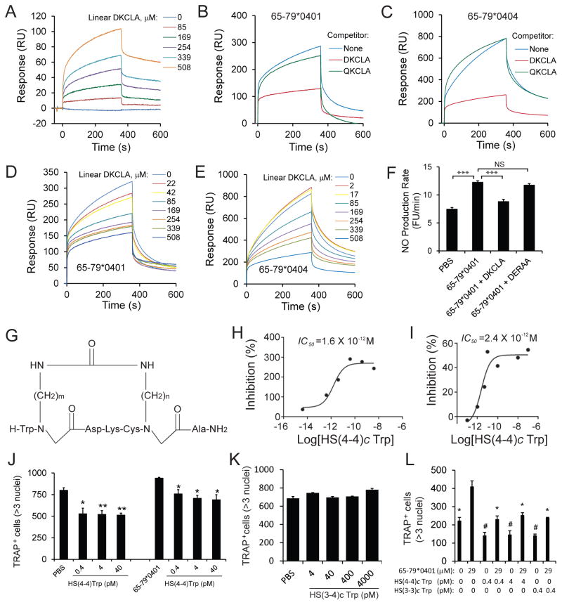
Methods: A library of backbone-cyclized peptidomimetic compounds, all carrying an identical core DKCLA sequence, was synthesized. The ability of these compounds to inhibit SE-activated signaling and OC differentiation was tested in vitro. The effect on disease severity and OC-mediated bone damage was studied by weekly intraperitoneal administration of the compounds to DBA/1 mice with CIA.
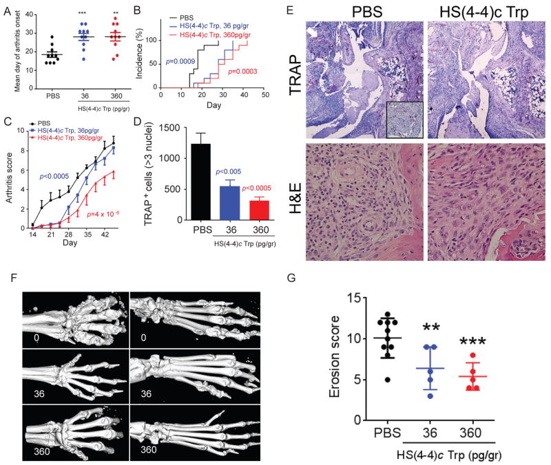
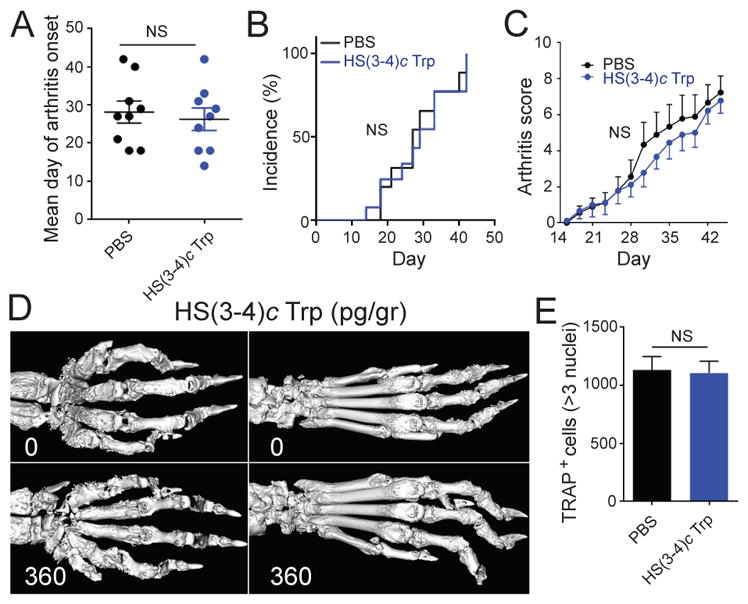
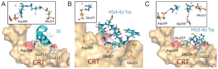
Results: Two members of the peptidomimetics library were found to have SE-antagonistic effects and antiosteoclast differentiation effects at picomolar concentrations in vitro. The lead mimetic compound, designated HS(4-4)c Trp, potently ameliorated arthritis and bone damage in vivo when administered in picogram doses to mice with CIA. Another mimetic analog, designated HS(3-4)c Trp, was found to lack activity, both in vitro and in vivo. The differential activity of the 2 analogs depended on minor differences in their respective ring sizes and correlated with distinctive geometry when computationally docked to the SE binding site on calreticulin.
Conclusion: These findings identify calreticulin as a novel therapeutic target in erosive arthritis and provide sound rationale and early structure/activity relationships for future drug design.
© 2015, American College of Rheumatology.
Figures
Comment in
-
Pharmacotherapy: Going upstream: peptidomimetics block shared-epitope signalling.Nat Rev Rheumatol. 2015 Jun;11(6):320. doi: 10.1038/nrrheum.2015.64. Epub 2015 May 5. Nat Rev Rheumatol. 2015. PMID: 25939417 No abstract available.
References
-
- Gregersen PK, Silver J, Winchester RJ. The shared epitope hypothesis. An approach to understanding the molecular genetics of susceptibility to rheumatoid arthritis. Arthritis Rheum. 1987;30:1205–13. - PubMed
-
- Gonzalez-Gay MA, Garcia-Porrua C, Hajeer AH. Influence of human leukocyte antigen-DRB1 on the susceptibility and severity of rheumatoid arthritis. Semin Arthritis Rheum. 2002;31:355–60. - PubMed
-
- Mattey DL, Hassell AB, Dawes PT, Cheung NT, Poulton KV, Thomson W, et al. Independent association of rheumatoid factor and the HLA-DRB1 shared epitope with radiographic outcome in rheumatoid arthritis. Arthritis Rheum. 2001;44:1529–33. - PubMed
-
- Plant MJ, Jones PW, Saklatvala J, Ollier WE, Dawes PT. Patterns of radiological progression in early rheumatoid arthritis: results of an 8 year prospective study. J Rheumatol. 1998;25:417–26. - PubMed
-
- Weyand CM, Goronzy JJ. Disease mechanisms in rheumatoid arthritis: gene dosage effect of HLA-DR haplotypes. J Lab Clin Med. 1994;124:335–8. - PubMed
Publication types
MeSH terms
Substances
Grants and funding
LinkOut - more resources
Full Text Sources
Other Literature Sources
Medical
Research Materials
