. 2015 Apr 28;11(4):539-50.
doi: 10.1016/j.celrep.2015.03.047. Epub 2015 Apr 16.

Cleavage-independent HIV-1 Env trimers engineered as soluble native spike mimetics for vaccine design

Affiliations

Cleavage-independent HIV-1 Env trimers engineered as soluble native spike mimetics for vaccine design

Shailendra Kumar Sharma et al. Cell Rep. .

Abstract

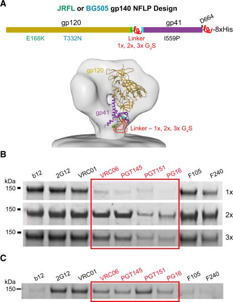
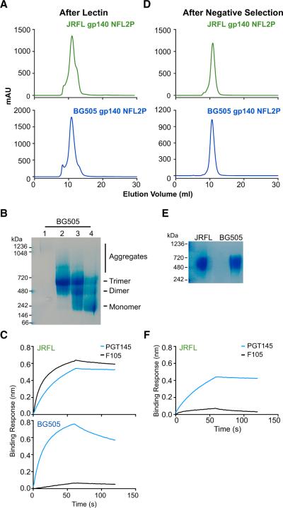
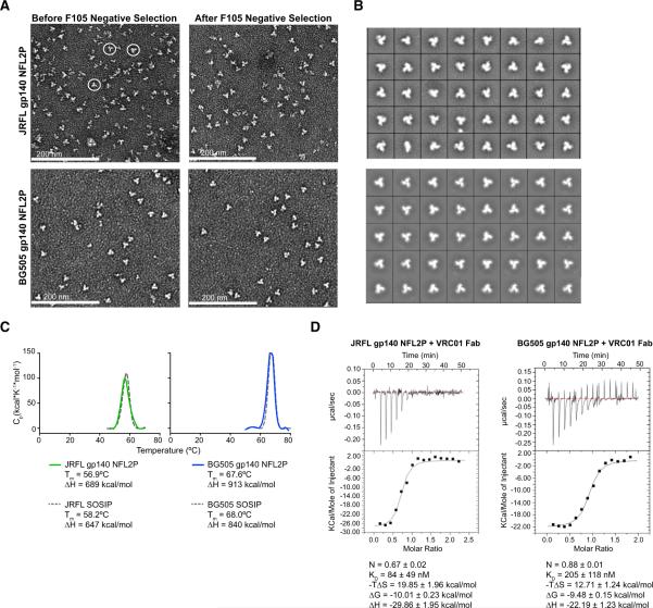
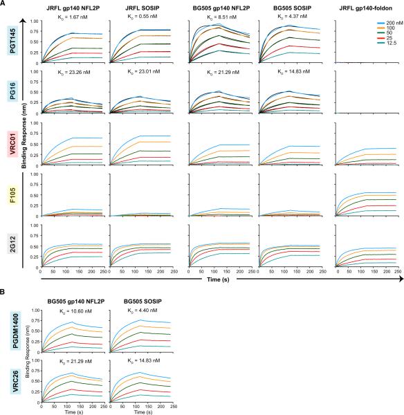
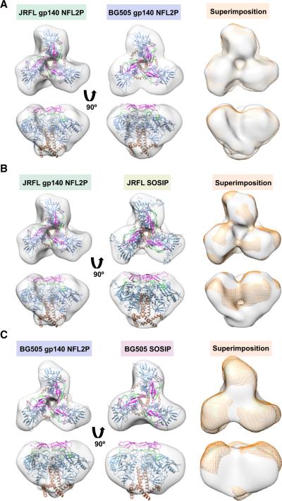
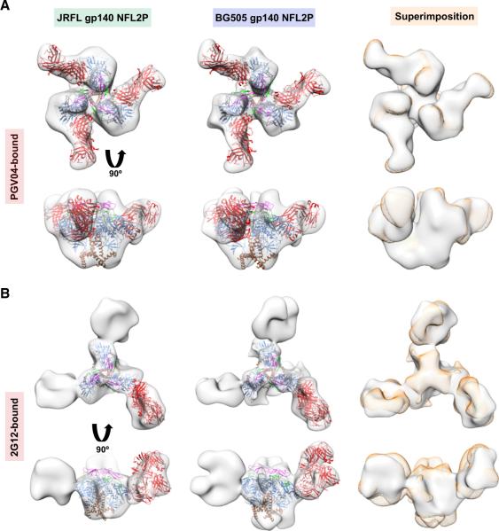
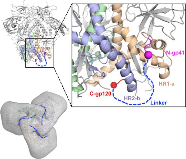
Viral glycoproteins mediate entry by pH-activated or receptor-engaged activation and exist in metastable pre-fusogenic states that may be stabilized by directed rational design. As recently reported, the conformationally fixed HIV-1 envelope glycoprotein (Env) trimers in the pre-fusion state (SOSIP) display molecular homogeneity and structural integrity at relatively high levels of resolution. However, the SOSIPs necessitate full Env precursor cleavage, which requires endogenous furin overexpression. Here, we developed an alternative strategy using flexible peptide covalent linkage of Env subdomains to produce soluble, homogeneous, and cleavage-independent Env mimics, called native flexibly linked (NFL) trimers, as vaccine candidates. This simplified design avoids the need for furin co-expression and, in one case, antibody affinity purification to accelerate trimer scale-up for preclinical and clinical applications. We have successfully translated the NFL design to multiple HIV-1 subtypes, establishing the potential to become a general method of producing native-like, well-ordered Env trimers for HIV-1 or other viruses.


Figures

Figure 1
Figure 1. NFL trimer design and linker length screening
(A) Top, schematic representing the NFL trimer design. The NFL glycoprotein trimers contain an N-terminal CD5 leader sequence followed by gp120 covalently linked to gp41 by 1, 2 or 3 copies of G4S flexible peptide linkers. The C-terminus of gp41 has one copy of the G4S linker followed by a His8 tag. Both proteins were designed with and without the I559P substitution. JRFL gp140 NFLs possess an additional substitution of E168K for PG9/PG16 recognition, while BG505 gp140 NFLs have a T332N mutation for the N332 glycan supersite. Bottom, model of gp120 (in golden rod with C-terminus in green) linked to gp41 (in purple with N-terminus in turquoise) by different linker lengths (red dashed lines) in the context of the viral spike; one protomer is shown for clarity. (B) JRFL gp140 NFLP with 1-3x G4S linkers were screened by IP with selected mAbs, Trimer-specific bNAbs are highlighted in red. (C). IP of BG505 gp140 NFL2P from crude cell supernatant by selected mAbs (trimer-specific mAbs highlighted in red). Note that the IP band intensity for non-NAb F105 is relatively light compared to those of the bNAbs, suggesting the majority of expressed protein is well-ordered trimers. See also Figure S1.
Figure 2
Figure 2. Purification of JRFL and BG505 gp140 NFL2P
(A). SEC profile of lectin purified JRFL (top) and BG505 (bottom) gp140 NFL2Ps. (B) Blue native gel analysis of select fractions from SEC of BG505 gp140 NFL2P. Lane 1) first peak, aggregates, 2) second peak, trimer, 3-4) third peak, dimer/monomer. (C) PGT145 and F105 binding profile of JRFL (top) and BG505 (bottom) gp140 NFL2P from SEC purified trimer peak by BLI. (D) SEC profile of JRFL (top) and BG505 (bottom) gp140 NFL2P after negative selection. (E) Blue native gel analysis of JRFL and BG505 gp140 NFL2P after negative selection. (F) PGT145 and F105 binding profile of JRFL gp140 NFL2P after negative selection. See also Figure S1C.
Figure 3
Figure 3. Negative stain EM analysis and thermal stability of purified JRFL and BG505 gp140 NFL2P trimers
(A) Negative stain EM micrographs of JRFL (top) and BG505 (bottom) gp140 NFL2P before (left) and after (right) negative selection. For JRFL, negative selection enriched for well-ordered trimers, whereas, no noticeable difference was observed for BG505 before and after negative selection. White scale bars, 200 nm. (B) 2D class averages of JRFL (top) and BG505 (bottom) gp140 NFL2P. (C) DSC melting profiles comparing JRFL (left) and BG505 (right) gp140 NFL2P trimers to respective SOSIP trimers. (D) The raw data (top) and binding isotherms (bottom) from ITC measuring the binding of JRFL (left) and BG505 (right) gp140 NFL2P to VRC01 Fab. See also Figure S2.
Figure 4
Figure 4. Antigenic profiles of JRFL and BG505 trimers by BLI
Anti-human Fc sensors were used to capture the mAbs and the trimers were used as an analyte at various concentrations (200-12.5 nM). The black curves for PG145 and PG16 in (A) and PGDM1400 and VRC26 in (B) depict theoretical Langmuir fits generated by 1:1 binding kinetics. Trimer-specific bNAbs (PGT145, PG16, PGDM1400, and VRC26); CD4bs-directed bNAb, VRC01; glycan-directed bNAb, 2G12; non-Nab, F105. See also Figure S3 and Table S1.
Figure 5
Figure 5. EM 3D reconstructions of unliganded JFRL and BG505 gp140 NFL2P compared with JRFL and BG505 SOSIP
(A) Top and side views of unliganded JRFL (left) and BG505 (middle) gp140 NFL2P 3D reconstruction EM densities in gray. The BG505 SOSIP crystal structure (PDB 3J5M) is fitted within (gp120 in blue, V1V2 in magenta, V3 in green and gp41 in brown). On the right, BG505 uncleaved trimer EM density (orange) is superimposed on the JRFL uncleaved trimer EM density (gray). (B) Top and side views of unliganded JRFL gp140 NFL2P and JRFL SOSIP in gray with BG505 SOSIP crystal structure fitted within as in (A). To the right, JRFL gp140 NFL2P EM density (orange) is superimposed on JRFL SOSIP EM density (gray). (C) Top and side views of unliganded BG505 gp140 NFL2P and BG505 SOSIP in gray with BG505 SOSIP crystal structure fitted within as above. On the right, BG505 gp140 NFL2P EM density (orange) is superimposed on BG505 SOSIP EM density (gray). See also Figures S4-5 and Table S2.
Figure 6
Figure 6. EM 3D reconstructions of PGV04-liganded and 2G12-liganded JRFL and BG505 gp140 NFL2P trimers
(A) Top and side views of the PGV04-liganded uncleaved trimers (in gray) with the PGV04-bound gp140 structure (PDB 3J5M; gp120 in blue, V1V2 in magenta, V3 in green, gp41 in brown and PGV04 in red) fitted within the EM density. (B) Top and side views of the 2G12-liganded uncleaved trimers with the 2G12-bound gp120 core crystal structure (PDB 1OP5; gp120 in blue, V1V2 in magenta, V3 in green, gp41 in brown and 2G12 in red) fitted in the EM density. On the right of each panel are top and side views of the BG505 uncleaved trimer EM density (in orange) superimposed on the JRFL uncleaved trimer EM density (in gray). See also Figures S4-5 and Table S2.
Figure 7
Figure 7. Model of the gp120-gp41 flexible linker using the BG505 SOSIP structure
Top left, high-resolution crystal structure of the PGT122- and 35O22-liganded BG505 SOSIP trimer (PDB 4TVP) with gp120 subunits colored in gray and gp41 subunits colored in green, violet and wheat, respectively (Fabs not shown for clarity). Top right, enlargement of the gp41-gp120 associated regions within the trimer. The blue dash line represents the flexible linker joining the last residue of the gp120 C-terminus (in red) to the first residue (visible in this structure) of the gp41 fusion peptide (in magenta). Bottom left, BG505 SOSIP crystal structure fitted within the EM density of the JRFL gp140 NFL2P trimer. Blue dotted lines represent the three flexible linkers on the surface EM contours, joining each of the three subunits that comprise the soluble NFL Env trimer.

References

    1. Binley JM, Sanders RW, Clas B, Schuelke N, Master A, Guo Y, Kajumo F, Anselma DJ, Maddon PJ, Olson WC, et al. A recombinant human immunodeficiency virus type 1 envelope glycoprotein complex stabilized by an intermolecular disulfide bond between the gp120 and gp41 subunits is an antigenic mimic of the trimeric virion- associated structure. J. Virol. 2000;74:627–643. - PMC - PubMed
    1. Blattner C, Lee JH, Sliepen K, Derking R, Falkowska E, de la Pena AT, Cupo A, Julien JP, van Gils M, Lee PS, et al. Structural delineation of a quaternary, cleavage-dependent epitope at the gp41-gp120 interface on intact HIV-1 Env trimers. Immunity. 2014;40:669–680. - PMC - PubMed
    1. Doria-Rose NA, Schramm CA, Gorman J, Moore PL, Bhiman JN, DeKosky BJ, Ernandes MJ, Georgiev IS, Kim HJ, Pancera M, et al. Developmental pathway for potent V1V2-directed HIV-neutralizing antibodies. Nature. 2014;509:55–62. - PMC - PubMed
    1. Falkowska E, Le KM, Ramos A, Doores KJ, Lee JH, Blattner C, Ramirez A, Derking R, van Gils MJ, Liang CH, et al. Broadly neutralizing HIV antibodies define a glycan-dependent epitope on the prefusion conformation of gp41 on cleaved envelope trimers. Immunity. 2014;40:657–668. - PMC - PubMed
    1. Forsell MN, Schief WR, Wyatt RT. Immunogenicity of HIV-1 envelope glycoprotein oligomers. Curr. Opin. HIV AIDS. 2009;4:380–387. - PubMed

Publication types

MeSH terms

Substances