Review
. 2015 Jun;185(6):1505-17.
doi: 10.1016/j.ajpath.2015.03.003. Epub 2015 Apr 17.

Pathogenesis and immunobiology of brucellosis: review of Brucella-host interactions

Affiliations
Review

Pathogenesis and immunobiology of brucellosis: review of Brucella-host interactions

Paul de Figueiredo et al. Am J Pathol. 2015 Jun.

Abstract

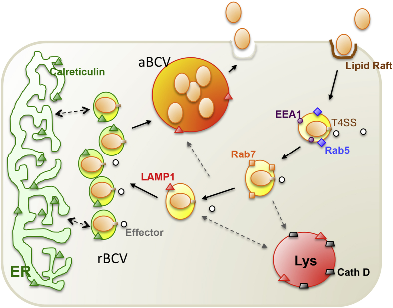
This review of Brucella-host interactions and immunobiology discusses recent discoveries as the basis for pathogenesis-informed rationales to prevent or treat brucellosis. Brucella spp., as animal pathogens, cause human brucellosis, a zoonosis that results in worldwide economic losses, human morbidity, and poverty. Although Brucella spp. infect humans as an incidental host, 500,000 new human infections occur annually, and no patient-friendly treatments or approved human vaccines are reported. Brucellae display strong tissue tropism for lymphoreticular and reproductive systems with an intracellular lifestyle that limits exposure to innate and adaptive immune responses, sequesters the organism from the effects of antibiotics, and drives clinical disease manifestations and pathology. Stealthy brucellae exploit strategies to establish infection, including i) evasion of intracellular destruction by restricting fusion of type IV secretion system-dependent Brucella-containing vacuoles with lysosomal compartments, ii) inhibition of apoptosis of infected mononuclear cells, and iii) prevention of dendritic cell maturation, antigen presentation, and activation of naive T cells, pathogenesis lessons that may be informative for other intracellular pathogens. Data sets of next-generation sequences of Brucella and host time-series global expression fused with proteomics and metabolomics data from in vitro and in vivo experiments now inform interactive cellular pathways and gene regulatory networks enabling full-scale systems biology analysis. The newly identified effector proteins of Brucella may represent targets for improved, safer brucellosis vaccines and therapeutics.


Figures

Figure 1
Figure 1
Hepatic and vertebral histopathology of human brucellosis caused by Brucella melitensis. A: Percutaneous liver biopsy. Mild nonspecific lymphocytic periportal hepatitis (arrow); stained with H&E. B: Percutaneous liver biopsy, culture positive for Brucella melitensis. Early-stage hepatic microgranuloma formation (arrow); stained with H&E. C: Guided needle core biopsy of vertebral body and epidural abscess, culture positive for Brucella melitensis. Lymphohistiocytic discitis osteomyelitis with dense cellular aggregates (arrow); stained with Diff-quik. Panels A and B are reproduced from Young et al with permission from Elsevier. Panel C was provided by Drs. Supriya Narasimhan and Michael L. Deftos (Santa Clara Valley Medical Center, San Jose, CA). Original magnification, ×40. H&E, hematoxylin and eosin.
Figure 2
Figure 2
Working model of Brucella intracellular trafficking in macrophage cells. Plasma membrane-associated lipid rafts mediate the internalization of smooth Brucella into macrophage cells. As the BCV matures, it sequentially associates with markers for early (EEA1, purple circle; Rab5, blue diamond) and late (Rab7, orange square) endosomes. The biogenesis and trafficking of BCVs is regulated by bacterial effector proteins (white circles), which are secreted through the Brucella T4SS. BCVs that contain virulent organisms do not fuse with lysosomes (cathepsin D, gray trapezoid), although transient association with LAMP1-positive membranes (orange triangles) is observed. The pathogen replicates in tight rBCVs that are decorated with calreticulin (green triangle), a marker for the ER. At a later point after infection (48 to 72 hours), the pathogen is observed in LAMP1-positive aBCVs that also contain LAMP1. The biogenesis of aBCVs depends on the activities of a subset of autophagy proteins, including ULK1 (not shown) and Beclin1 (not shown). Finally, the pathogen is released from the cell through lytic or nonlytic (shown) mechanisms. aBCV, autophagic Brucella-containing vacuole; BCV, Brucella-containing vacuole; Beclin1, coiled-coil myosin-like BCL2-interacting protein; EEA1, early endosome antigen 1; ER, endoplasmic reticulum; LAMP1, lysosome-associated membrane protein 1; rBCV, replicative Brucella-containing vacuole; T4SS, type IV secretion system; ULK1, Unc-51-like kinase 1.
Figure 3
Figure 3
Scheme of in vivo and/or in vitro systems biology analysis of host and Brucella interactive pathogenesis. Experimental pathology includes the collection and omics (transcriptomics, proteomics, metabolomics, etc.) data from both mammalian host and Brucella samples from an in vivo time series of Brucella infection of a target organ (eg, Peyer patch, lung, spleen, liver) in a natural target animal (eg, cattle, sheep, goat, pig, nonhuman primate). The resulting omics (eg, transcriptomics, proteomics, metabolomics) data sets are fused and bioinformatically analyzed for known and computed structural modeling of predicted host–pathogen protein–protein interactions to develop an in silico interactome structure learning model. Proteins are inferred from genes if not directly measured. Pairs of predicted candidate Brucella–host protein–protein mechanistic genes from interactive pathways are phenotyped in vitro in standardized gentamicin killing assays by using specific deletion mutants of Brucella, siRNA knockdown of host genes, and confocal microscopy. Brucella–host protein–protein pairs with positive in vitro phenotypes are phenotyped in vivo, and high confidence positive candidate protein pairs undergo pull-down analysis and quantitative selected reaction monitoring mass spectrometry for further confirmation of Brucella–host protein–protein interactions in Brucella pathogenesis. Blue lines and arrows indicate flow of in vivo data and results from the host, and brown lines and arrows indicate flow of data and results from Brucella into in silico analysis to identify and model domain A from a host protein predicted to interact with domain B of Brucella.
Figure 4
Figure 4
Brucella is bipolar. Brucella both inhibits and promotes a proinflammatory immune response. TLR4 signaling during infection is restricted by the presence of elongated fatty acid chains that reduce the toxicity of the LPS (blue studs on Brucella surface) and by blocking downstream IKK phosphorylation via MyD88 binding (blue squares) with Brucella TIR-containing proteins, BtpA and BtpB (red squares), leading to enhanced polyubiquitination and degradation of MAL. However, the T4SS (VjbR-controlled expression) effector VceC (red oval) stimulates an innate immune response via interaction with BiP, an ER molecular chaperone (green squares) to release and phosphorylate IRE1 to promote mRNA splicing of XBP1 and activation of UPR. IRE1 phosphorylation also promotes the proinflammatory response via the release of NF-κB from the complex with IκBα (maroon crescent). The critical distinction between these two pathways may reside in the timing of activation. Inhibiting the host early or MyD88-mediated response may promote acquisition of a replicative niche, whereas the delayed T4SS-mediated VceC effector (VjbR-controlled expression) response may enhance the spread of the organism. Solid arrows indicate Brucella-mediated activation, whereas dotted arrows indicate Brucella-mediated inhibition. ER, endoplasmic reticulum; IκBα, inhibitor of κB protein α; Iκκ, IκB kinase; IRE1, inositol-requiring enzyme 1; LPS, lipopolysaccharide; MAL, MyD88-adaptor like; MYd88, myeloid differentiation response gene 88; T4SS, type IV secretion system; TIRAP, Toll–IL-1 receptor domain-containing adaptor protein; TLR4, Toll-like receptor 4; TNF-α, tumor necrosis factor α; UPR, unfolded protein response; XBP1, X-box binding protein 1.

References

    1. Pappas G., Panagopoulou P., Christou L., Akritidis N. Brucella as a biological weapon. Cell Mol Life Sci. 2006;63:2229–2236. - PMC - PubMed
    1. Young E.J., Hasanjani Roushan M.R., Shafae S., Genta R.M., Taylor S.L. Liver histology of acute brucellosis caused by Brucella melitensis. Hum Pathol. 2014;45:2023–2028. - PubMed
    1. Dean A.S., Crump L., Greter H., Hattendorf J., Schelling E., Zinsstag J. Clinical manifestations of human brucellosis: a systematic review and meta-analysis. PLoS Negl Trop Dis. 2012;6:e1929. - PMC - PubMed
    1. Martirosyan A., Gorvel J.P. Brucella evasion of adaptive immunity. Future Microbiol. 2013;8:147–154. - PubMed
    1. Baud D., Greub G. Intracellular bacteria and adverse pregnancy outcomes. Clin Microbiol Infect. 2011;17:1312–1322. - PubMed

Publication types