

Left ventricular diastolic reserve in patients with type 2 diabetes mellitus
- PMID: 25893102
- PMCID: PMC4395831
- DOI: 10.1136/openhrt-2014-000214
Left ventricular diastolic reserve in patients with type 2 diabetes mellitus
Abstract
Aims: Diastolic reserve is the ability of left ventricular filling pressures to remain normal with exercise. Impaired diastolic reserve may be an early sign of diabetic cardiomyopathy. We aimed to determine whether diastolic reserve differs in type 2 diabetes (DM) compared with non-DM, and to identify clinical, anthropological, metabolic and resting echocardiographic correlates of impaired diastolic reserve in patients with DM.
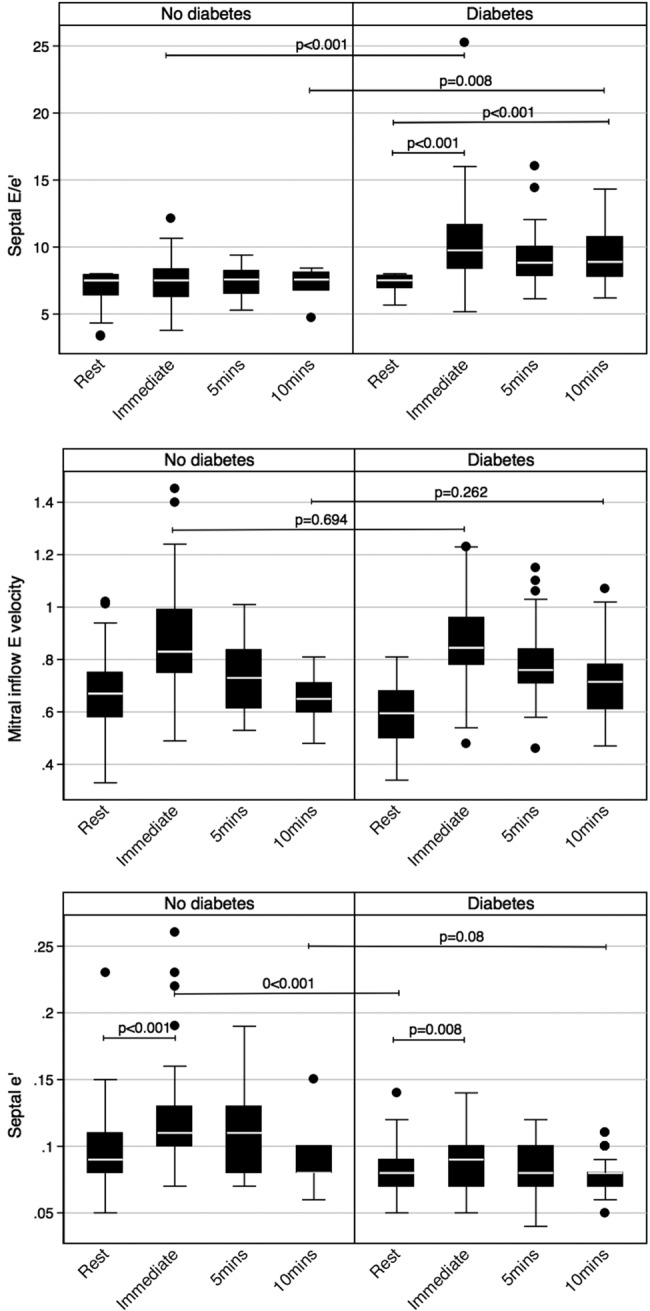
Methods and results: 237 patients (aged 53±11 years, 133 DM, ejection fraction 68±9%) underwent rest and exercise echocardiography. Mitral E and septal e' were measured at rest, immediately post, and 10 min into recovery. Analysis of covariance (ANCOVA) and binary regression with continuous outcomes were used to model e' and E/e' changes with exercise to identify impaired diastolic reserve defined as post-exercise E/e' ≥15. After adjusting for baseline differences, patients with DM immediately post-exercise had a lower septal e', a lower Δe' (1.2 vs 2.3 cm/s, p=0.006) and a higher Δ septal E/e' (1.7 vs 0.08, p<0.001) than patients without DM. In patients with normal resting E/e' of ≤8 (n=130), DM had a significantly higher post-exercise septal E/e' and a higher Δseptal E/e' (2.63 vs 0.50, p<0.001). E/e' in patients with DM remained significantly elevated up to 10 min post-exercise. Hypertension, longer duration of insulin therapy, poorer glycaemic control, worse renal function, larger left atrial volume and lower septal e' were independent correlates of impaired diastolic reserve in patients with DM.
Conclusions: Patients with DM have impaired diastolic reserve manifest as a blunted e' response with exercise, persisting into recovery. Clinical, anthropometric, metabolic and echocardiographic correlates of impaired diastolic reserve in patients with DM were identified. An impaired LV diastolic reserve may be the underlying pathophysiological mechanism in patients with DM with unexplained exertional dyspnoea and may allow earlier detection of DM cardiomyopathy.
Figures
Similar articles
-
Endothelial function and left ventricular diastolic functional reserve in type 2 diabetes mellitus.Open Heart. 2014 Aug 5;1(1):e000113. doi: 10.1136/openhrt-2014-000113. eCollection 2014. Open Heart. 2014. PMID: 25332819 Free PMC article.
-
Incremental value of left ventricular diastolic function reserve index for predicting exercise capacity in patients with hypertrophic cardiomyopathy.J Am Soc Echocardiogr. 2008 May;21(5):487-92. doi: 10.1016/j.echo.2007.08.041. Epub 2007 Sep 29. J Am Soc Echocardiogr. 2008. PMID: 17904804
-
Heterogeneous responses of systolic and diastolic left ventricular function to exercise in patients with heart failure and preserved ejection fraction.ESC Heart Fail. 2015 Sep;2(3):121-132. doi: 10.1002/ehf2.12049. Epub 2015 Aug 10. ESC Heart Fail. 2015. PMID: 27708854 Free PMC article.
-
Correlation with invasive left ventricular filling pressures and prognostic relevance of the echocardiographic diastolic parameters used in the 2016 ESC heart failure guidelines and in the 2016 ASE/EACVI recommendations: a systematic review in patients with heart failure with preserved ejection fraction.Eur J Heart Fail. 2018 Sep;20(9):1303-1311. doi: 10.1002/ejhf.1220. Epub 2018 Jun 7. Eur J Heart Fail. 2018. PMID: 29877602
-
Diastolic stress echocardiography: from basic principles to clinical applications.Heart. 2018 Nov;104(21):1739-1748. doi: 10.1136/heartjnl-2017-312323. Epub 2018 Jul 20. Heart. 2018. PMID: 30030333 Review.
Cited by
-
Abnormal left ventricular systolic reserve function detected by treadmill exercise stress echocardiography in asymptomatic type 2 diabetes.Front Cardiovasc Med. 2023 Oct 19;10:1253440. doi: 10.3389/fcvm.2023.1253440. eCollection 2023. Front Cardiovasc Med. 2023. PMID: 37928757 Free PMC article.
-
Improvement of continuous subcutaneous insulin infusion on patients with type 2 diabetes mellitus by 3-dimensional speckle tracking echocardiography.Int J Cardiovasc Imaging. 2018 Mar;34(3):379-384. doi: 10.1007/s10554-017-1245-5. Epub 2017 Sep 21. Int J Cardiovasc Imaging. 2018. PMID: 28936576
-
Mechanical loading reveals an intrinsic cardiomyocyte stiffness contribution to diastolic dysfunction in murine cardiometabolic disease.J Physiol. 2024 Dec;602(24):6705-6727. doi: 10.1113/JP286437. Epub 2024 Dec 4. J Physiol. 2024. PMID: 39629708 Free PMC article.
-
Effects of 1,25-dihydroxyvitamin D3 on pathological changes in rats with diabetic cardiomyopathy.Lipids Health Dis. 2017 Jun 8;16(1):109. doi: 10.1186/s12944-017-0498-2. Lipids Health Dis. 2017. PMID: 28595623 Free PMC article.
-
Vascular endothelial dysfunction, a major mediator in diabetic cardiomyopathy.Acta Pharmacol Sin. 2019 Jan;40(1):1-8. doi: 10.1038/s41401-018-0042-6. Epub 2018 Jun 4. Acta Pharmacol Sin. 2019. PMID: 29867137 Free PMC article. Review.
References
Publication types
LinkOut - more resources
Full Text Sources
Other Literature Sources