

Human GM3 Synthase Attenuates Taxol-Triggered Apoptosis Associated with Downregulation of Caspase-3 in Ovarian Cancer Cells
- PMID: 25893133
- PMCID: PMC4399708
- DOI: 10.4236/jct.2012.35065
Human GM3 Synthase Attenuates Taxol-Triggered Apoptosis Associated with Downregulation of Caspase-3 in Ovarian Cancer Cells
Abstract
Background: Taxol (paclitaxel) inhibits proliferation and induces apoptosis in a variety of cancer cells, but it also upregulates cytoprotective proteins and/or pathways that compromise its therapeutic efficacy.
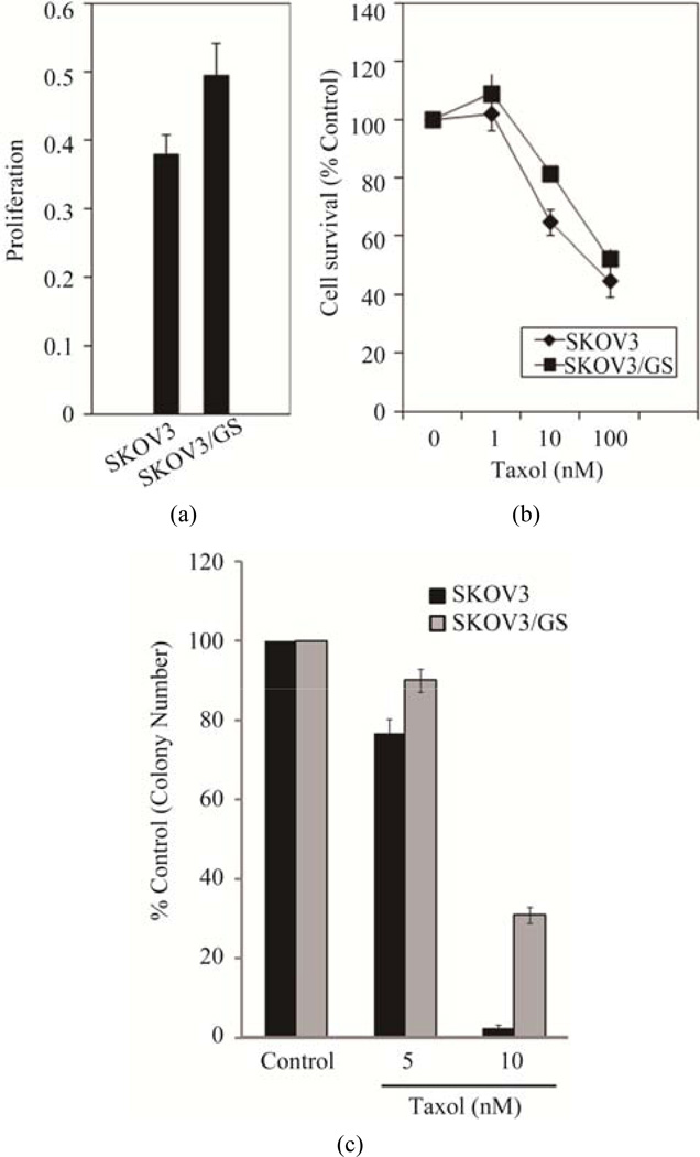
Materials and method: The roles of GM3 synthase (α2,3-sialyltransferase, ST3Gal V) in attenuating Taxol-induced apoptosis and triggering drug resistance were determined by cloning and overexpressing this enzyme in the SKOV3 human ovarian cancer cell line, treating SKOV3 and the transfectants (SKOV3/GS) with Taxol and determining apoptosis, cell survival, clonogenic ability, and caspase-3 activation.
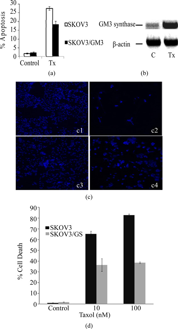
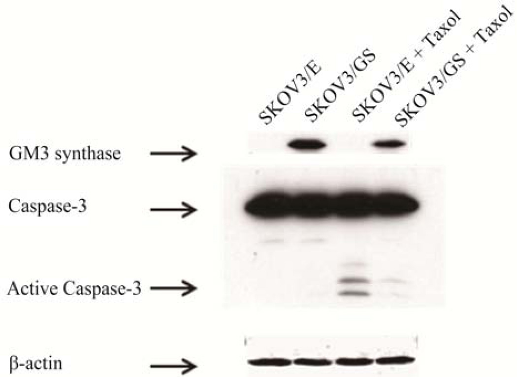
Results: In this report, we demonstrated that Taxol treatment resulted in apoptosis which was associated with caspase-3 activation. Taxol treatment upregulated the expression of human GM3 synthase, an enzyme that transfers a sialic acid to lactosylceramide. Moreover, we cloned the full-length GM3 synthase gene and showed for the first time that forced expression of GM3 synthase attenuated Taxol-induced apoptosis and increased resistance to Taxol in SKOV3 cells.
Conclusions: GM3 synthase overexpression inhibited Taxol-triggered caspase-3 activation, revealing that upregulation of GM3 synthase prevents apoptosis and hence reduces the efficacy of Taxol therapy.
Keywords: Apoptosis; Caspase-8; Drug Resistance; GM3 Synthase; Ovarian Cancer; Taxol.
Figures
References
-
- Daly M, Obrams GI. Epidemiology and Risk Assessment for Ovarian Cancer. Seminars in Oncology. 2008;25(3):255–264. - PubMed
-
- Crown J, O’Leary M, Ooi WS. Docetaxel and Paclitaxel in the Treatment of Breast Cancer: A Review of Clinical Experience. Oncologist. 2004;9(Suppl. 2):24–32. - PubMed
-
- Fields AL, Runowicz CD. Current Therapies in Ovarian Cancer. Cancer Investigation. 2003;21(1):148–156. - PubMed
-
- Jordan MA, Wilson L. Microtubules as a Target for Anticancer Drugs. Nature Reviews Cancer. 2004;4(4):253–265. - PubMed
-
- Liao PC, Lieu CH. Cell Cycle Specific Induction of Apoptosis and Necrosis by Paclitaxel in the Leukemic U937 Cells. Life Sciences. 2005;76(14):1623–1639. - PubMed
Grants and funding
LinkOut - more resources
Full Text Sources
Other Literature Sources
Research Materials