. 2014 Dec 30;1(1):e20.14.
doi: 10.1523/ENEURO.0006-14.2014.

Altered Brain Structure and Function Correlate with Disease Severity and Pain Catastrophizing in Migraine Patients

Affiliations

Altered Brain Structure and Function Correlate with Disease Severity and Pain Catastrophizing in Migraine Patients

Catherine S Hubbard et al. eNeuro. .

Abstract

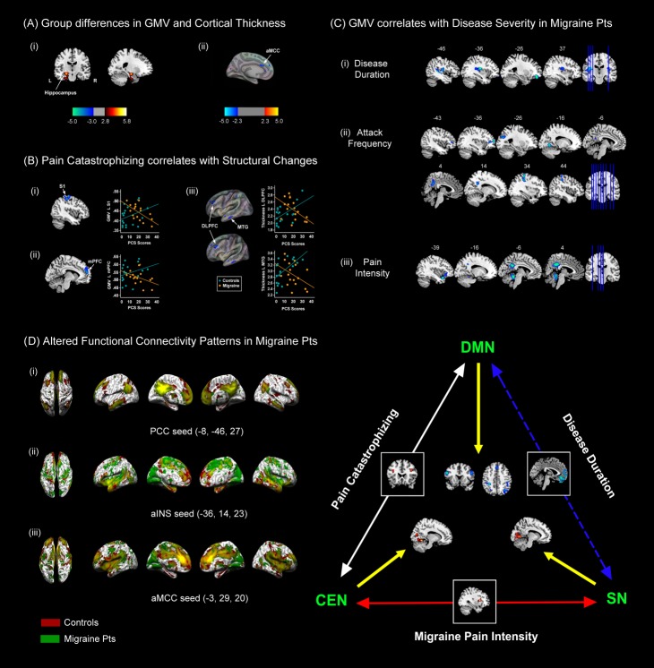
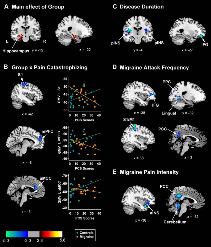
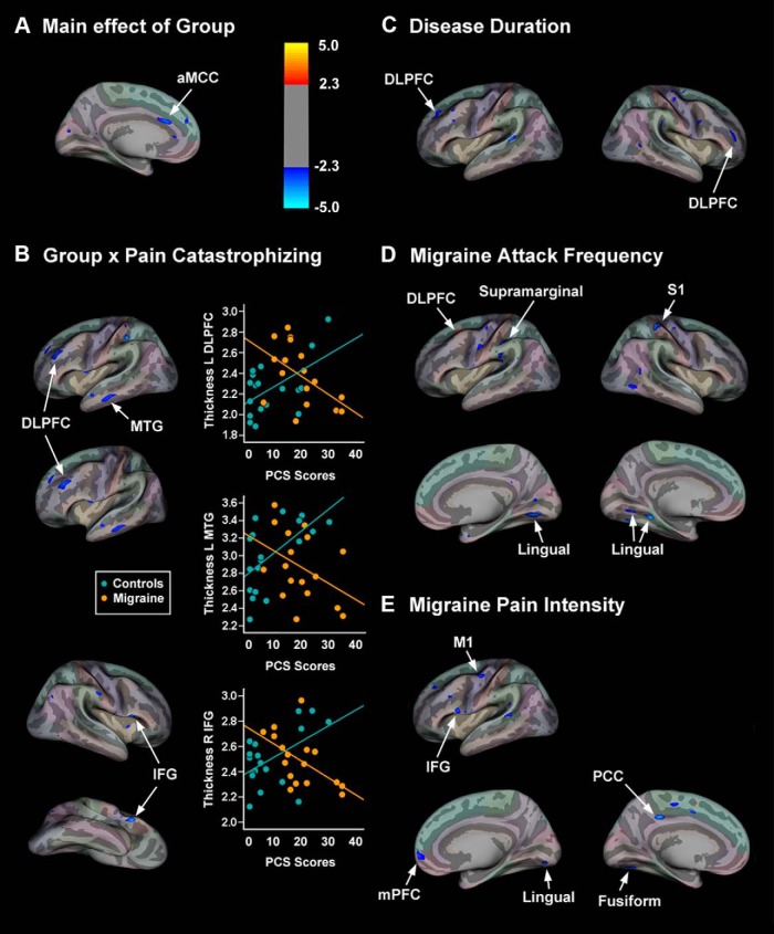
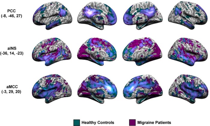
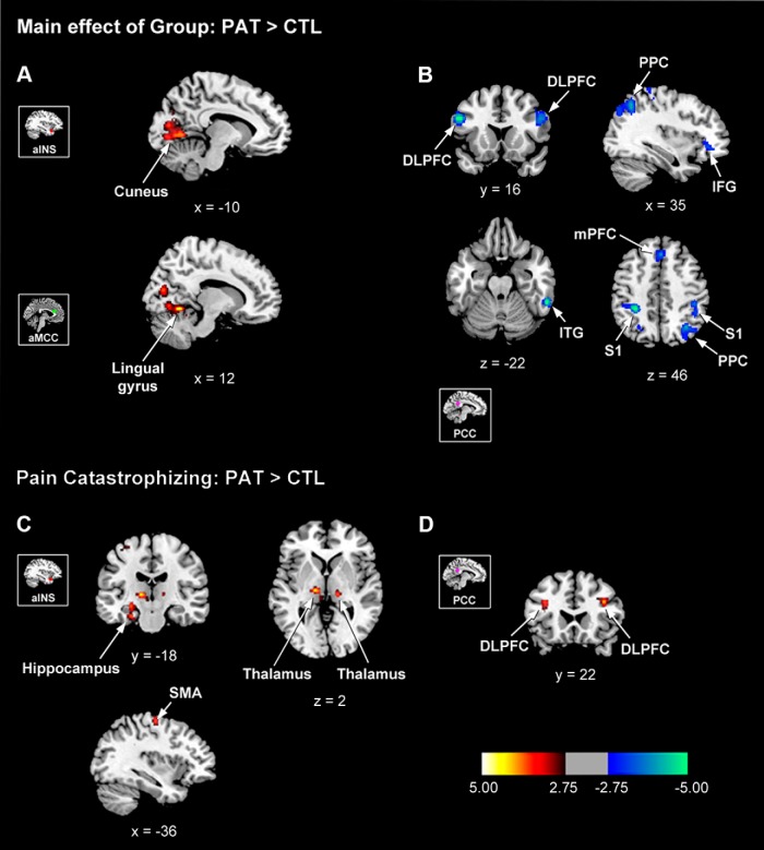
To investigate the neuroanatomical and functional brain changes in migraine patients relative to healthy controls, we used a combined analytical approach including voxel- and surface-based morphometry along with resting-state functional connectivity to determine whether areas showing structural alterations in patients also showed abnormal functional connectivity. Additionally, we wanted to assess whether these structural and functional changes were associated with group differences in pain catastrophizing and migraine-related disease variables in patients. We acquired T1-weighted anatomical and functional magnetic resonance imaging scans during rest in human subjects with a diagnosis of migraine and healthy controls. Structural analyses revealed greater left hippocampal gray matter volume and reduced cortical thickness in the left anterior midcingulate in patients compared with controls. We also observed negative associations between pain catastrophizing and migraine disease variables and gray matter in areas implicated in processing the sensory, affective, and cognitive aspects of pain in patients. Functional connectivity analyses showed that migraine patients displayed disrupted connectivity between default mode, salience, cognitive, visuospatial, and sensorimotor networks, which was associated with group differences in pain catastrophizing and migraine-related disease variables in patients. Together, our findings show widespread morphological and functional brain abnormalities in migraineurs in affective, cognitive, visual, and pain-related brain areas, which are associated with increased pain catastrophizing, disease chronicity, and severity of symptoms, suggesting that these structural and functional changes may be a consequence of repeated, long-term nociceptive signaling leading to increased pain sensitivity, mood disturbances, and maladaptive coping strategies to deal with unrelenting pain.

Keywords: chronic pain; gray matter; headache; intrinsic connectivity; neuroimaging; resting-state networks.


Conflict of interest statement

We thank the University of Maryland Magnetic Resonance Research Center, Dr. Rao Gullapalli and George Makris for their assistance with data collection. We would also like to express our gratitude to all the participants who took part in this study. The authors declare no competing financial interests.

Figures

Cover Figure
Cover Figure
Migraine patients (Pts) show widespread structural and functional brain changes that are associated with symptoms and increased pain catastrophizing A, Migraine patients showed (i) increased gray matter volume (GMV) in the left (L) hippocampus and (ii) decreased cortical thickness in the L anterior midcingulate cortex (aMCC) compared to healthy control subjects. B, Pain catastrophizing correlated with GMV reductions in the (i) L primary somatosensory cortex (S1) and (ii) L medial prefrontal cortex (mPFC), and cortical thinning in the (iii) L dorsolateral prefrontal cortex (DLPFC) and middle temporal gyrus (MTG) in migraine patients. C, GMV reductions correlated with (i) disease duration (ii), attack frequency, and (iii) migraine pain intensity in patients. D, Whole-brain overlay maps for migraine patients and healthy controls for the (i) L PCC, (ii) L aINS, and (iii) aMCC seed regions rendered onto inflated brains. Red represents resting-state functional connectivity for healthy controls and green represents the same maps in migraine patients. Yellow represents areas showing overlap in functional connectivity in controls and migraineurs. Images are thresholded at T = 4.5 (cluster extent = 25) for visualization purposes. The schematic illustrates the relationship between disease severity measures and pain catastrophizing and disruptions in functional connectivity between the default mode network (DMN), central executive network (CEN), and salience network (SN) in migraine patients. In patients, pain catastrophizing correlated with increased coupling between DMN and CEN nodes (PCC-DLPFC), whereas disease duration and migraine pain intensity correlated with SN-DMN network decoupling (aINS/aMCC-mPFC), and increased SN-CEN (aMCC-aINS) network coupling, respectively.
Figure 1.
Figure 1.
Statistical maps for the voxel-based morphometric analysis overlaid onto the averaged ch2bet brain template in MRIcron. A, Patients showed significant increased GMV in the left hippocampus, which extended into the parahippocampal gyrus, compared with controls. L, Left hemisphere; R, right hemisphere. B, Cortical areas showing significant group × pain catastrophizing interactions for GMV and corresponding scatter plots showing correlations between GMV and catastrophizing in migraine patients (orange circles) and healthy controls (blue circles). C−E, Regions showing significant reductions in GMV that correlated with longer disease durations (C), greater attack frequency (D), and migraine pain intensity in patients (E).
Figure 2.
Figure 2.
Statistical maps for the surface-based analysis rendered onto the inflated averaged brain template using Freesurfer’s QDEC graphical user interface software. A, Group differences in cortical thickness were found; migraine patients showed significant reductions in cortical thickness in the left aMCC compared with healthy controls. B, Cortical areas showing significant group × pain catastrophizing interactions for cortical thickness measures and corresponding scatter plots displaying correlations between cortical thickness and catastrophizing in migraine patients (orange circles) and healthy controls (blue circles). C−E, Cortical areas showing significant cortical thinning (blue) and thickening (red) in migraine patients that were associated with longer disease durations (C), greater migraine attack frequency (D), and migraine pain intensity in patients (E).
Figure 3.
Figure 3.
Whole-brain overlay maps for patients and controls for the left PCC, left aINS, and aMCC seed regions, rendered onto inflated brains in SPM8. Teal represents RS-FC for healthy controls and purple represents the same maps in migraine patients. Light violet represents areas showing overlap in RS-FC in controls and patients. Images are thresholded at T = 4.5 (cluster extent = 25) for visualization purposes. The PCC seed showed a connectivity map consistent with the DMN, whereas the aINS and aMCC seeds connectivity patterns were consistent with the SN.
Figure 4.
Figure 4.
Group differences in RS-FC for the aINS and aMCC seeds (SN) and PCC seed (DMN) overlaid onto the average ch2bet brain template in MRIcron. A, Patients showed significant enhancement in aINS and aMCC connectivity with the L cuneus and R lingual gyrus, respectively, and B, reduced connectivity between the PCC seed and bilateral DLPFC, S1, R PPC, R IFG, R ITG, and R mPFC. C, D, RS-FC group differences associated with pain catastrophizing for the aINS and PCC seeds. C, Patients high in catastrophizing showed enhanced RS-FC between the aINS and the L hippocampus, L SMA, and bilateral thalami. D, Significant increases in RS-FC were also identified between the PCC and the bilateral DLPFC in patients with greater pain catastrophizing scores. PAT, Patients; CTL, controls.

References

    1. Archer DP, Roth SH (1997) Pharmacodynamics of thiopentone: nocifensive reflex threshold changes correlate with hippocampal electroencephalography. Br J Anaesth 79:744-749. - PubMed
    1. Ashburner J (2007) A fast diffeomorphic image registration algorithm. Neuroimage 38:95-113. 10.1016/j.neuroimage.2007.07.007 - DOI - PubMed
    1. Ashburner J, Friston KJ (2000) Voxel-based morphometry: the methods. Neuroimage 11:805-821. 10.1006/nimg.2000.0582 - DOI - PubMed
    1. Ashburner J, Friston KJ (2001) Why voxel-based morphometry should be used. Neuroimage 14:1238-1243. 10.1006/nimg.2001.0961 - DOI - PubMed
    1. Baliki MN, Geha PY, Apkarian AV, Chialvo DR (2008) Beyond feeling: chronic pain hurts the brain, disrupting the default-mode network dynamics. J Neurosci 28:1398-1403. 10.1523/JNEUROSCI.4123-07.2008 - DOI - PMC - PubMed

LinkOut - more resources