

Correlation of Brain Atrophy, Disability, and Spinal Cord Atrophy in a Murine Model of Multiple Sclerosis
- PMID: 25893491
- PMCID: PMC4506196
- DOI: 10.1111/jon.12250
Correlation of Brain Atrophy, Disability, and Spinal Cord Atrophy in a Murine Model of Multiple Sclerosis
Abstract
Background: Disability progression in multiple sclerosis (MS) remains incompletely understood. Unlike lesional measures, central nervous system atrophy has a strong correlation with disability. Theiler's murine encephalomyelitis virus infection in SJL/J mice is an established model of progressive MS. We utilized in vivo MRI to quantify brain and spinal cord atrophy in this model and analyzed the temporal relationship between atrophy and disability.
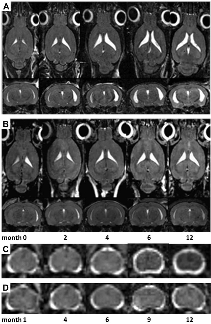
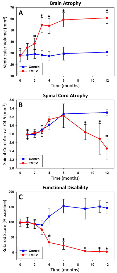
Methods: Infected and control mice were followed for 12 months. Disability was assessed periodically using rotarod assay. Volumetric MRI datasets were acquired at 7 Tesla. Ventricular volume and C4-5 spinal cord cross-sectional area measurements were performed using Analyze 10.
Results: At 3 months, brain atrophy reached statistical significance (P = .005). In contrast, disability did not differ until 4 months post-infection (P = .0005). Cord atrophy reached significance by 9 months (P = 0.009). By 12 months, brain atrophy resulted in 111.8% increased ventricular volume (P = .00003), while spinal cord cross-sectional area was 25.6% reduced (P = .001) among cases.
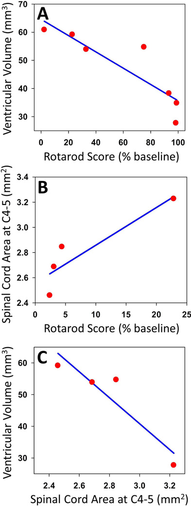
Conclusions: Our results suggest that significant brain atrophy precedes and predicts the development of disability, while spinal cord atrophy occurs late and correlates with severe disability. The observed temporal relationship establishes a framework for mechanisms of disability progression and enables further investigations of their underlying substrate.
Keywords: Multiple sclerosis; atrophy; disability; magnetic resonance imaging.
Copyright © 2015 by the American Society of Neuroimaging.
Conflict of interest statement
Figures
References
-
- Prineas JW. Pathology of multiple sclerosis. In: Cook SD, editor. Handbook of multiple sclerosis. 3rd. New York: Marcel Dekker; 2001. pp. 289–324.
-
- Stadelmann C, Albert M, Wegner C, Bruck W. Cortical pathology in multiple sclerosis. Curr Opin Neurol. 2008;21:229–234. - PubMed
-
- Trapp BD, Nave KA. Multiple sclerosis: an immune or neurodegenerative disorder? Annu Rev Neurosci. 2008;31:247–269. - PubMed
-
- De Stefano N, Matthews PM, Filippi M, et al. Evidence of early cortical atrophy in MS: relevance to white matter changes and disability. Neurology. 2003;60:1157–1162. - PubMed
-
- Kalkers NF, Ameziane N, Bot JC, Minneboo A, Polman CH, Barkhof F. Longitudinal brain volume measurement in multiple sclerosis: rate of brain atrophy is independent of the disease subtype. Arch Neurol. 2002;59:1572–1576. - PubMed
Publication types
MeSH terms
Grants and funding
LinkOut - more resources
Full Text Sources
Other Literature Sources
Medical
Miscellaneous
