. 2016 Jan;127(1):91-102.
doi: 10.1007/s11120-015-0141-x. Epub 2015 Apr 19.

Resolving the contribution of the uncoupled phycobilisomes to cyanobacterial pulse-amplitude modulated (PAM) fluorometry signals

Affiliations

Resolving the contribution of the uncoupled phycobilisomes to cyanobacterial pulse-amplitude modulated (PAM) fluorometry signals

Alonso M Acuña et al. Photosynth Res. 2016 Jan.

Abstract

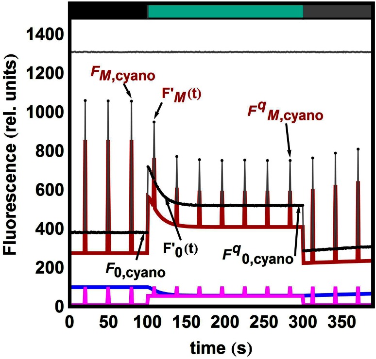
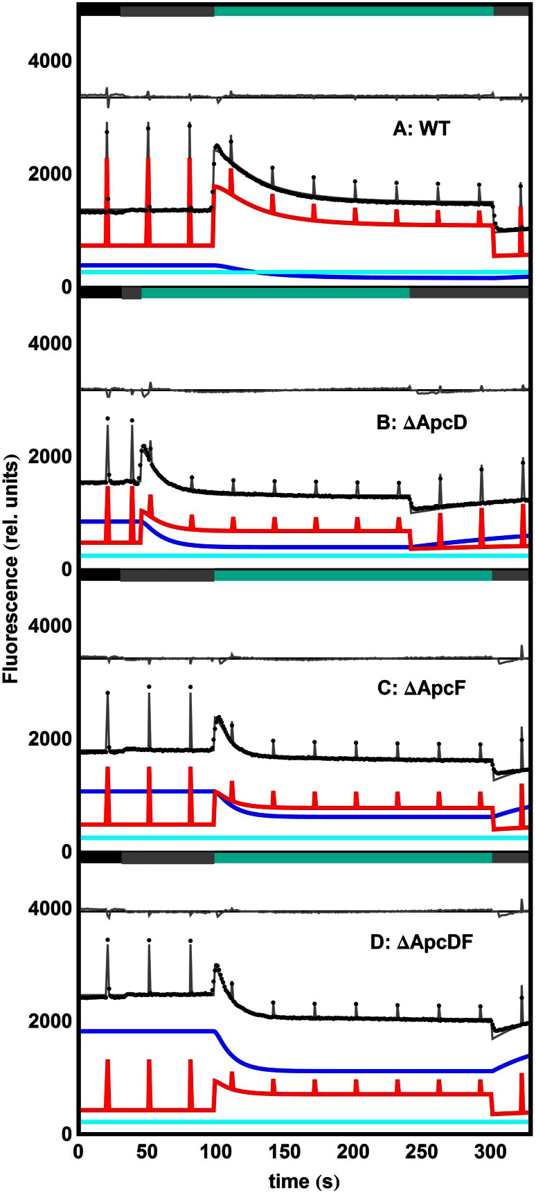
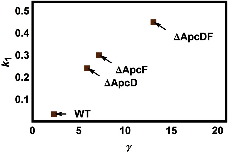
Pulse-amplitude modulated (PAM) fluorometry is extensively used to characterize photosynthetic organisms on the slow time-scale (1-1000 s). The saturation pulse method allows determination of the quantum yields of maximal (F(M)) and minimal fluorescence (F(0)), parameters related to the activity of the photosynthetic apparatus. Also, when the sample undergoes a certain light treatment during the measurement, the fluorescence quantum yields of the unquenched and the quenched states can be determined. In the case of cyanobacteria, however, the recorded fluorescence does not exclusively stem from the chlorophyll a in photosystem II (PSII). The phycobilins, the pigments of the cyanobacterial light-harvesting complexes, the phycobilisomes (PB), also contribute to the PAM signal, and therefore, F(0) and F(M) are no longer related to PSII only. We present a functional model that takes into account the presence of several fluorescent species whose concentrations can be resolved provided their fluorescence quantum yields are known. Data analysis of PAM measurements on in vivo cells of our model organism Synechocystis PCC6803 is discussed. Three different components are found necessary to fit the data: uncoupled PB (PB(free)), PB-PSII complexes, and free PSI. The free PSII contribution was negligible. The PB(free) contribution substantially increased in the mutants that lack the core terminal emitter subunits allophycocyanin D or allophycocyanin F. A positive correlation was found between the amount of PB(free) and the rate constants describing the binding of the activated orange carotenoid protein to PB, responsible for non-photochemical quenching.

Keywords: Cyanobacteria; Fluorescence quantum yield; Non-photochemical quenching; Phycobilisome; Pulse-amplitude modulated (PAM) fluorometry.


Figures

Fig. 1
Fig. 1
Emissive spectra of Chl a and PB in vivo. The spectral overlap within the detection region of typical PAM setups is shown in red
Fig. 2
Fig. 2
The different emissive species giving rise to a cyanobacterial PAM signal [see Eq. (3)] are characterized by their respective quantum yields in different light-acclimated states. Phycobilisomes are represented as six blue rods attached to three grey cylinders as the core. Two rectangular units represent an open (dark green) or closed (red) PSII-dimer. PBs can be bound to a PS or not. The index j indicates whether the emissive species is an uncoupled PB (a j = PB) a single PB–PSII complex (PPc2) (b j = PPc2). j = PB can still result in two different states if, for instance, a minimal amount of light (black bar) or high-light (blue–green bar) is shone onto the sample: unquenched (c l = u) or quenched (d l = q). The index k indicates whether the PSII is open (e k = o) or, in case a saturation pulse fully reduces the PQ pool, closed (f k = c)
Fig. 3
Fig. 3
Simulated data with a pulse every 20 s and noise level of 0.01. At time t = 100 s, strong blue-green light is turned on (indicated by the colour bars on the top). Its power is such that a fraction c 0 of the RCs are closed and the OCPo → OCPr conversion takes place with κ I = 0.09 s−1. The amount of OCPr formed is [OCPr] = 0.5 and it binds to PB with k 1 = 0.30 s−1. The FRP detaches the OCP bound to the PB with k 2 = 0.003 s−1. A fluorescence recovery region has been added: at time t = 300 s, the NPQ-inducing light is turned off. All RCs re-open and the OCPo → OCPr conversion stops (κ I = 0). The action of FRP can be resolved better and k 2 can be estimated (k 2 = 0.00306 ± 0.00007 s−1). For all estimated parameters, see Table S2. Black dots simulated data points; red PPc2 contribution; blue PBfree contribution; magenta PSIIfree contribution; grey sum of the three contributions. Residuals are shown on top with an offset of 1300
Fig. 4
Fig. 4
Fit of experimental PAM measurements performed on whole cells of WT Synechocystis (a) and mutants thereof: ∆ApcD (b), ∆ApcF (c), ∆ApcDF (d). Colour bars on the top of each panel indicate the light regime: dark (black), 30 µE (grey) and 1400 µE (turquoise). All estimated parameters are collated in Table 2. Black dots experimental data points; red PPc2 contribution; blue PBfree contribution; cyan PSIfree contribution; grey sum of all contributions. Residuals (grey) are shown in each panel with an offset
Fig. 5
Fig. 5
Correlation between the estimated parameters γ and k 1 in the different samples

References

    1. Adir N. Elucidation of the molecular structures of components of the phycobilisome: reconstructing a giant. Photosynth Res. 2005;85:15–32. doi: 10.1007/s11120-004-2143-y. - DOI - PubMed
    1. Arteni AA, Ajlani G, Boekema EJ. Structural organisation of phycobilisomes from Synechocystis sp strain PCC6803 and their interaction with the membrane. Biochim Biophys Acta. 2009;1787:272–279. doi: 10.1016/j.bbabio.2009.01.009. - DOI - PubMed
    1. Baker NR. Chlorophyll fluorescence: a probe of photosynthesis in vivo. Annu Rev Plant Biol. 2008;59:89–113. doi: 10.1146/annurev.arplant.59.032607.092759. - DOI - PubMed
    1. Boulay C, Wilson A, D’Haene S, Kirilovsky D. Identification of a protein required for recovery of full antenna capacity in OCP-related photoprotective mechanism in cyanobacteria. Proc Natl Acad Sci USA. 2010;107:11620–11625. doi: 10.1073/pnas.1002912107. - DOI - PMC - PubMed
    1. Bryant DA. Cyanobacterial phycobilisomes: progress toward complete structural and functional analysis via molecular genetics. In: Bogorad L, Vasil IK, editors. The photosynthetic apparatus: molecular biology and operation. San Diego: Academic Press; 1991. pp. 257–300.

Publication types

MeSH terms

LinkOut - more resources