. 2015 Jun;47(6):654-60.
doi: 10.1038/ng.3279. Epub 2015 Apr 20.

COPA mutations impair ER-Golgi transport and cause hereditary autoimmune-mediated lung disease and arthritis

Affiliations

COPA mutations impair ER-Golgi transport and cause hereditary autoimmune-mediated lung disease and arthritis

Levi B Watkin et al. Nat Genet. 2015 Jun.

Abstract

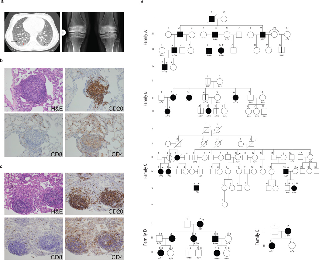
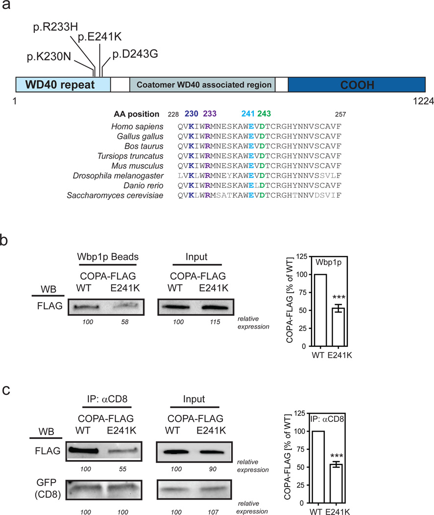
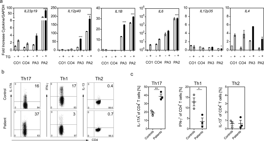
Unbiased genetic studies have uncovered surprising molecular mechanisms in human cellular immunity and autoimmunity. We performed whole-exome sequencing and targeted sequencing in five families with an apparent mendelian syndrome of autoimmunity characterized by high-titer autoantibodies, inflammatory arthritis and interstitial lung disease. We identified four unique deleterious variants in the COPA gene (encoding coatomer subunit α) affecting the same functional domain. Hypothesizing that mutant COPA leads to defective intracellular transport via coat protein complex I (COPI), we show that COPA variants impair binding to proteins targeted for retrograde Golgi-to-ER transport. Additionally, expression of mutant COPA results in ER stress and the upregulation of cytokines priming for a T helper type 17 (TH17) response. Patient-derived CD4(+) T cells also demonstrate significant skewing toward a TH17 phenotype that is implicated in autoimmunity. Our findings uncover an unexpected molecular link between a vesicular transport protein and a syndrome of autoimmunity manifested by lung and joint disease.


Figures

Figure 1
Figure 1. Genetic segregation of ILD and arthritis with COPA mutations
(a) On left, a chest CT scan of patient B.III.1 reveals basilar ground glass opacities (circle), reticulations (red arrow) and cystic formations (black arrow). Right panel is a bilateral knee radiograph from A.III.6 prior to restorative surgery. The radiograph shows generalized osteopenia with pathologic fracture of the distal femur. There is bilateral avascular necrosis of the metaphysis and diaphysis of the distal femur and proximal tibia with slackening of the condyles and articular cartilage destruction. (b,c) Lung biopsy sections from patients A.III.6, and B.III.1 stained with hematoxylin and eosin show cellular interstitial infiltrates and germinal center formation while immunohistochemical analysis demonstrates presence of CD20+ B cells and CD4+ and CD8+ T cells. (d) The pedigrees for Families A-E are shown with the presence of a wild type (+) or mutant (m) COPA allele. Family A (c.G698A) shows incomplete penetrance with one unaffected mutant carrier over four generations. Family B (c.A728G) shows incomplete penetrance of severe disease in females affected over three generations. Family C (c.G721A) shows incomplete penetrance of severe disease and segregation of the mutation within affected patients over six generations. For Family D (c.G690T) there is an unaffected mutation carrier who is 1 year old, which is below the age of onset. Family E (c.G698A) shows complete penetrance over two generations. Square=male; circle=female; black filled=affected with severe disease; unfilled=unaffected; /=deceased; *=submitted for WES; vertical lines=unaffected carrier. Roman numerals represent the generation within the family and Arabic numerals designate individuals within the generation.
Figure 2
Figure 2. Binding of mutant COPA to dilysine motifs is impaired
(a) Schematic of the COPA protein and its domains is shown. The amino acid position of the mutations identified and the conserved sequence homology at those sites in distantly related eukaryotic organisms are indicated. (b) Binding of mutant COPA to a dilysine motif was examined by taking either FLAG-tagged wild-type or mutant E241K COPA and incubating it with Wbp1p peptide-coupled agarose beads. The protein-bead complexes were analyzed by western blot (WB) using an anti-FLAG antibody. Numbers indicate the relative expression in percent of wild-type COPA. Statistical analysis of 6 independent experiments is shown. ***P<0.001 (c) Binding of mutant COPA to a dilysine motif was also assessed within T-Rex-293 cells stably expressing FLAG-tagged wild-type or mutant COPA by transiently transfecting cells with an expression construct for GFP-CD8-KKTN. Cell lysates were precipitated with anti-CD8 and immunoprecipitates were assayed for FLAG-COPA expression and GFP-CD8 enrichment. Numbers indicate the relative expression in percent of wild-type COPA or GFP-CD8 expressed in WT COPA cells. Statistical analysis of 5 independent experiments is shown. ***P<0.001.
Figure 3
Figure 3. Mutant COPA leads to an increase in ER stress
(a) Immunohistochemistry stain for the ER stress component BiP in lung biopsy samples from patients with E241K, D243G, or R233H mutant COPA is shown. Biopsies from individuals not carrying a COPA mutation were used as a control. Hematoxylin was used for nuclear counterstain. Scale bars indicate 40µm. Enlarged inserts emphasize BiP staining in lung epithelial cells. (b) Quantitative PCR (qPCR) analysis of BiP expression in untreated (white bars; −) B lymphoblastoid cell-lines (BLCL) and treated (striped, gray and black bars; +) BLCL after stimulation with thapsigargin. Left graph indicates the results from 4 controls (CO1 (N=12), CO2 (N=3), CO3 (N=4), CO4 (N=9); CO3 is a related control of Family D, D.III.6, and CO4 of Family C, C.III.10). All results were normalized to values of untreated control 1. Data shown are from a healthy carrier (HC1, C.III.2, N=9) and 2 patients (PA1-2, C.IV.15 (N=12), C.V.1 (N=8)) of the E241K COPA family C, a healthy carrier (HC2, B.III.4, N=5) and one patient (PA4, B.III.3, N=5) of the D243G COPA family B, and 2 patients (PA 5–6, A.II.5 (N=5), A.III.6 (N=4)) of the R233H family A. One way ANOVA followed by Dunnett's Multiple Comparison Test was used to compare thapsigargin-treated experimental groups with thapsigargin-treated control 1 as a reference. Mean and SEM of pooled data from 2–4 independent experiments are shown. *P<0.05, **P<0.01, ***P<0.001. (c) COPA knockdown in HEK 293 cells. Left graph demonstrates greater than 90% reduction in COPA mRNA expression levels after treatment with COPA siRNA compared to cells treated with control siRNA. Right graph shows the induction of ER stress measured by BiP expression after COPA knockdown. Mean and SEM of pooled data from 3 independent experiments are shown (N=11). Statistical comparisons were made using Student’s unpaired t test with Welch's correction. **P<0.01, ***P<0.001. (d) HEK 293 cells were transfected with wild-type COPA (N=17) or mutant E241K (N=14), D243G (N=12), or R233H (N=12) COPA expression constructs. The graph shows BiP expression 48h after transfection as assessed by qPCR. Cells transfected with mutant COPA showed significantly increased BiP levels when normalized to cells transfected with wild-type COPA. Mean and SEM are shown for each column. One-way ANOVA followed by Dunnett's Multiple Comparison Test was use to compare experimental groups against wild-type transfected HEK293 cells as the control column. Results shown are pooled data from 3 independent experiments. *P<0.05, **P<0.01, ***P<0.001. CO denotes control, HC healthy carrier, PA patient, TG thapsigargin, WT wild-type.
Figure 4
Figure 4. Patients with mutant COPA demonstrate an increase in Th17 priming cytokines in BLCLs and an increase in Th17 cells
(a) Cytokine expression was measured in BLCLs from CO1, CO4 (C.III.10), PA2 (C.V.1) and PA3 (C.IV.2) either untreated (white bars; −) or treated (grey, black bars; +) with TG. Samples were analyzed via qPCR for mRNA levels of IL23p19, IL12p40, IL12p35, IL4, IL1β, and IL6. Shown are normalized mRNA levels against the average value for healthy control 1 (CO1) in the absence of stimulation. Errors bars indicate the standard error for triplicates in the assay. Results are representative of 3 independent experiments. Nothing indicated P>0.05; *P<0.05; ***P<0.001. (b,c) CD4+ T cells from three COPA patients with stable disease (C.IV.1 and C.IV.2 were on mycophenolate; C.IV.15 was on no treatment) and three age-matched controls were analyzed for intracellular cytokines IL-17A, IFN-, and IL-13 after stimulation with PMA and Ionomycin. (b) Representative FACS plots for IL-17A, IFN-, and IL-13-producing CD4+ T cells are shown. Plots are gated on living CD3+/CD4+ T cells. (c) Graphs demonstrating the frequency of Th1, Th2 and Th17 cells in patients and controls. The frequency of IL-17A-secreting Th17 cells is significantly increased in patients compared to controls (patients mean 39% vs. control mean 19% **P<0.01; N=3) whereas patients show a significant decrease in IFN--producing Th1 cells (patient mean 4% vs. control mean 13% *P<0.05; N=3). No significant difference was found in the frequency of IL-13-secreting Th2 cells. Student’s unpaired t test was used for statistical evaluation.

Similar articles

Cited by

  • The Trinity of cGAS, TLR9, and ALRs Guardians of the Cellular Galaxy Against Host-Derived Self-DNA.
    Kumar V. Kumar V. Front Immunol. 2021 Feb 11;11:624597. doi: 10.3389/fimmu.2020.624597. eCollection 2020. Front Immunol. 2021. PMID: 33643304 Free PMC article. Review.
  • Innovations in Childhood Interstitial and Diffuse Lung Disease.
    Casey A, Fiorino EK, Wambach J. Casey A, et al. Clin Chest Med. 2024 Sep;45(3):695-715. doi: 10.1016/j.ccm.2024.04.002. Clin Chest Med. 2024. PMID: 39069332 Free PMC article. Review.
  • EMC10 homozygous variant identified in a family with global developmental delay, mild intellectual disability, and speech delay.
    Umair M, Ballow M, Asiri A, Alyafee Y, Al Tuwaijri A, Alhamoudi KM, Aloraini T, Abdelhakim M, Althagafi AT, Kafkas S, Alsubaie L, Alrifai MT, Hoehndorf R, Alfares A, Alfadhel M. Umair M, et al. Clin Genet. 2020 Dec;98(6):555-561. doi: 10.1111/cge.13842. Epub 2020 Sep 15. Clin Genet. 2020. PMID: 32869858 Free PMC article.
  • Primary immunodeficiency diseases: Genomic approaches delineate heterogeneous Mendelian disorders.
    Stray-Pedersen A, Sorte HS, Samarakoon P, Gambin T, Chinn IK, Coban Akdemir ZH, Erichsen HC, Forbes LR, Gu S, Yuan B, Jhangiani SN, Muzny DM, Rødningen OK, Sheng Y, Nicholas SK, Noroski LM, Seeborg FO, Davis CM, Canter DL, Mace EM, Vece TJ, Allen CE, Abhyankar HA, Boone PM, Beck CR, Wiszniewski W, Fevang B, Aukrust P, Tjønnfjord GE, Gedde-Dahl T, Hjorth-Hansen H, Dybedal I, Nordøy I, Jørgensen SF, Abrahamsen TG, Øverland T, Bechensteen AG, Skogen V, Osnes LTN, Kulseth MA, Prescott TE, Rustad CF, Heimdal KR, Belmont JW, Rider NL, Chinen J, Cao TN, Smith EA, Caldirola MS, Bezrodnik L, Lugo Reyes SO, Espinosa Rosales FJ, Guerrero-Cursaru ND, Pedroza LA, Poli CM, Franco JL, Trujillo Vargas CM, Aldave Becerra JC, Wright N, Issekutz TB, Issekutz AC, Abbott J, Caldwell JW, Bayer DK, Chan AY, Aiuti A, Cancrini C, Holmberg E, West C, Burstedt M, Karaca E, Yesil G, Artac H, Bayram Y, Atik MM, Eldomery MK, Ehlayel MS, Jolles S, Flatø B, Bertuch AA, Hanson IC, Zhang VW, Wong LJ, Hu J, Walkiewicz M, Yang Y, Eng CM, Boerwinkle E, Gibbs RA, Shearer WT, Lyle R, Orange JS, Lupski JR. Stray-Pedersen A, et al. J Allergy Clin Immunol. 2017 Jan;139(1):232-245. doi: 10.1016/j.jaci.2016.05.042. Epub 2016 Jul 16. J Allergy Clin Immunol. 2017. PMID: 27577878 Free PMC article.
  • Coatomer-associated protein subunit alpha syndrome: abnormal trafficking between the Golgi complex and the endoplasmic reticulum.
    Perrin J, Roux N, Maurier F. Perrin J, et al. BMJ Case Rep. 2020 Nov 2;13(11):e231553. doi: 10.1136/bcr-2019-231553. BMJ Case Rep. 2020. PMID: 33139352 Free PMC article.

References

    1. Zenewicz LA, Abraham C, Flavell RA, Cho JH. Unraveling the genetics of autoimmunity. Cell. 2010;140:791–797. - PMC - PubMed
    1. Letourneur F, et al. Coatomer is essential for retrieval of dilysine-tagged proteins to the endoplasmic reticulum. Cell. 1994;79:1199–1207. - PubMed
    1. Eugster A, Frigerio G, Dale M, Duden R. COP I domains required for coatomer integrity, and novel interactions with ARF and ARF-GAP. EMBO J. 2000;19:3905–3917. - PMC - PubMed
    1. Popoff V, Adolf F, Brügger B, Wieland F. COPI budding within the Golgi stack. Cold Spring Harbor Perspectives in Biology. 2011;3:a005231. - PMC - PubMed
    1. Leipe J, et al. Role of Th17 cells in human autoimmune arthritis. Arthritis Rheum. 2010;62:2876–2885. - PubMed

Publication types

MeSH terms

Substances