Bridging the gap between traditional cell cultures and bioreactors applied in regenerative medicine: practical experiences with the MINUSHEET perfusion culture system
- PMID: 25894791
- PMCID: PMC4754254
- DOI: 10.1007/s10616-015-9873-x
Bridging the gap between traditional cell cultures and bioreactors applied in regenerative medicine: practical experiences with the MINUSHEET perfusion culture system
Abstract
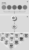
To meet specific requirements of developing tissues urgently needed in tissue engineering, biomaterial research and drug toxicity testing, a versatile perfusion culture system was developed. First an individual biomaterial is selected and then mounted in a MINUSHEET(®) tissue carrier. After sterilization the assembly is transferred by fine forceps to a 24 well culture plate for seeding cells or mounting tissue on it. To support spatial (3D) development a carrier can be placed in various types of perfusion culture containers. In the basic version a constant flow of culture medium provides contained tissue with always fresh nutrition and respiratory gas. For example, epithelia can be transferred to a gradient container, where they are exposed to different fluids at the luminal and basal side. To observe development of tissue under the microscope, in a different type of container a transparent lid and base are integrated. Finally, stem/progenitor cells are incubated in a container filled by an artificial interstitium to support spatial development. In the past years the described system was applied in numerous own and external investigations. To present an actual overview of resulting experimental data, the present paper was written.
Keywords: 3D culture; Biomaterial testing; Biomedicine; Bioreactor; Cell culture; Perfusion culture; Tissue carrier; Tissue engineering.
Figures


Similar articles
-
Supportive development of functional tissues for biomedical research using the MINUSHEET® perfusion system.Clin Transl Med. 2012 Oct 5;1(1):22. doi: 10.1186/2001-1326-1-22. Clin Transl Med. 2012. PMID: 23369669 Free PMC article.
-
A modular culture system for the generation of multiple specialized tissues.Biomaterials. 2010 Apr;31(11):2945-54. doi: 10.1016/j.biomaterials.2009.12.048. Epub 2010 Jan 22. Biomaterials. 2010. PMID: 20096452
-
Technical and theoretical considerations about gradient perfusion culture for epithelia used in tissue engineering, biomaterial testing and pharmaceutical research.Biomed Mater. 2007 Jun;2(2):R1-R11. doi: 10.1088/1748-6041/2/2/R01. Epub 2007 Mar 7. Biomed Mater. 2007. PMID: 18458434 Review.
-
Renal epithelia in long term gradient culture for biomaterial testing and tissue engineering.Biomed Mater Eng. 2005;15(1-2):51-63. Biomed Mater Eng. 2005. PMID: 15623930
-
Spaceflight bioreactor studies of cells and tissues.Adv Space Biol Med. 2002;8:177-95. doi: 10.1016/s1569-2574(02)08019-x. Adv Space Biol Med. 2002. PMID: 12951697 Review.
Cited by
-
Apical Medium Flow Influences the Morphology and Physiology of Human Proximal Tubular Cells in a Microphysiological System.Bioengineering (Basel). 2022 Sep 30;9(10):516. doi: 10.3390/bioengineering9100516. Bioengineering (Basel). 2022. PMID: 36290484 Free PMC article.
-
In vitro biomimetic platforms featuring a perfusion system and 3D spheroid culture promote the construction of tissue-engineered corneal endothelial layers.Sci Rep. 2017 Apr 10;7(1):777. doi: 10.1038/s41598-017-00914-1. Sci Rep. 2017. PMID: 28396609 Free PMC article.
References
-
- Aigner J, Kloth S, Kubitza M, Kashgarian M, Dermietzel R, Minuth WW (1994) Maturation of renal collecting duct cells in vivo and under perifusion culture. Epithel Cell Biol 3:70–78 - PubMed
-
- Aigner J, Kloth S, Jennings ML, Minuth WW (1995) Transitional differentiation patterns of principal and intercalated cells during renal collecting duct development. Epithel Cell Biol 4:121–130 - PubMed
-
- Anders JO, Mollenhauer J, Beberhold A, Kinne RW, Venbrocks RA. Gelatin-based haemostyptic Spongostan as a possible three-dimensional scaffold for a chondrocyte matrix? An experimental study with bovine chondrocytes. J Bone Joint Surg Br. 2009;91:409–416. doi: 10.1302/0301-620X.91B3.20869. - DOI - PubMed
LinkOut - more resources
Full Text Sources
Other Literature Sources
Miscellaneous

