. 2015 May;21(5):467-475.
doi: 10.1038/nm.3842. Epub 2015 Apr 20.

Homeostatic regulation of T cell trafficking by a B cell-derived peptide is impaired in autoimmune and chronic inflammatory disease

Affiliations

Homeostatic regulation of T cell trafficking by a B cell-derived peptide is impaired in autoimmune and chronic inflammatory disease

Myriam Chimen et al. Nat Med. 2015 May.

Abstract

During an inflammatory response, lymphocyte recruitment into tissue must be tightly controlled because dysregulated trafficking contributes to the pathogenesis of chronic disease. Here we show that during inflammation and in response to adiponectin, B cells tonically inhibit T cell trafficking by secreting a peptide (PEPITEM) proteolytically derived from 14.3.3 zeta delta (14.3.3.ζδ) protein. PEPITEM binds cadherin-15 on endothelial cells, promoting synthesis and release of sphingosine-1 phosphate, which inhibits trafficking of T cells without affecting recruitment of other leukocytes. Expression of adiponectin receptors on B cells and adiponectin-induced PEPITEM secretion wanes with age, implying immune senescence of the pathway. Additionally, these changes are evident in individuals with type 1 diabetes or rheumatoid arthritis, and circulating PEPITEM in patient serum is reduced compared to that of healthy age-matched donors. In both diseases, tonic inhibition of T cell trafficking across inflamed endothelium is lost. Control of patient T cell trafficking is re-established by treatment with exogenous PEPITEM. Moreover, in animal models of peritonitis, hepatic ischemia-reperfusion injury, Salmonella infection, uveitis and Sjögren's syndrome, PEPITEM reduced T cell recruitment into inflamed tissues.


Figures

Figure 1
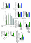
Figure 1. T cell migration across endothelial cells is regulated by a soluble agent released from B cells stimulated with adiponectin
(a-c) The effects of adiponectin (0–15 μg/ml) on T cell migration across TNF-α&IFN-γ treated (a) HUVECs under static (n=3–7) or (b) flow conditions (n=6), or (c), HDMEC under static conditions (n=3–4). (d) Representative plots of Adiponectin receptor-1 and -2 (AdipoR1 and 2) expression on T cells (CD3+) and B cells (CD19+) measured by flow-cytometry. (e, f) Percentage of adiponectin receptor positive cells on lymphocyte subpopulations (measured by flow-cytometry), n=6–13. (g) Transmigration of T cells following B cell depletion, reconstitution of depleted preparations with B cells or B cell supernatant or inhibition B cell secretion by Brefeldin A (10 μg/ml), n=3–7. Data are from at least 3 independent experiments using 3 donors for both PBL and HUVEC/HDMEC and are mean±s.e.m. NS: not significant, * P≤0.05, ** P≤0.01, *** P≤0.001 by (a) linear regression (b-c, g) paired t-test compared to untreated (no adiponectin) control or (e, f) Dunnett post-test compared to B cells.
Figure 2
Figure 2. PEPITEM inhibits T cell transmigration by binding to Cadherin-15 on endothelial cells
(a) MS/MS analysis of ion m/z 774.8 with the predicted sequence for PEPITEM, data representative of n=3. (b) Amino acid sequence of 14.3.3.ζδ with PEPITEM highlighted in blue. (c) The effect of PEPITEM (0–10 ng/ml), scrambled control peptide or irrelevant peptides (proinsulin chain A and Tetanus toxoid peptide at 10 ng/ml) on PBL transmigration, n=3–4. (d) The effect of PEPITEM on the transmigration of pre-sorted leukocyte populations. Data normalized to scrambled control peptide, n=3–6. NA= no adhesion. (e, f) The effects of control or cadherin-15 (CDH15) specific siRNA on (e) the mRNA expression of CDH15 in HDMECs (n=6) and (f) on T cell transmigration across HDMECs (n=6). Data normalized to (e) unstimulated or (f) untreated and no siRNA control. (g-h) The effects of control or CDH15 specific siRNA on the protein expression of CDH15 determined by (g) Western Blot or (h) confocal microscopy in HDMEC and skeletal muscle cells (SkMC). Scale bars: 30 μm. Data are mean±s.e.m. * P≤0.05, ** P≤0.01, *** P≤0.001 compared to (c) no adiponectin/PEPITEM control, (d) scrambled control and (e, f) unstimulated control or no siRNA by Dunnett post-test.
Figure 3
Figure 3. PEPITEM induces the S1P release from endothelial cells which inhibits T cell migration
(a, b) The effects of an S1PR antagonist (10 μM) on T cell migration in the presence or absence of (a) adiponectin (n=3–5) or (b) PEPITEM (n=3–7). (c) The effects on T cell transmigration of adding S1P to B cell depleted PBL n=3–6. (d, e) The effects of (d) SPHK1 specific inhibitor (5 μM) or (e) SPHK1/2 inhibitor (5 μM) on T cell transmigration in the presence of PEPITEM, n=3. (f) The expression of SPHK1 and SPHK2 mRNA in endothelial cells, n=7–8. (g) The effect of SPNS2 specific siRNA on T cell transmigration in the presence of PEPITEM, n=4. (h, i) The expression of S1PR1 on memory T cells (CD3+CD45RO+) on (h) stimulated endothelial cells (n=3) and (i) on plated ICAM-1 stimulated with CXCL10 (n=6). (j) The effects of S1P on the expression of the LFA-1 activation epitope (KIM127) on ICAM-1 adherent memory T cells (CD3+CD45RO+) stimulated with CXCL10, n=4. Data are mean±s.e.m and (g-j) normalized to control. * P≤0.05, ** P≤0.01, *** P≤0.001 compared to untreated control by ANOVA and (a-g) Dunnett compared to (a-b, d-e, g) untreated control or (c) B cell depletion no adiponectin control or to (f) unstimulated (0) control or (j) Bonferroni post-test or (h) paired t-test on raw data or (i) Wilcoxon signed rank test.
Figure 4
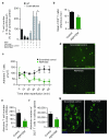
Figure 4. PEPITEM inhibits T cell migration in vivo
(a) T cells recruited into the peritoneum of BALB/c B cell-deficient mice after induction of peritonitis using zymosan and treatment with PEPITEM or scrambled peptide. All data was normalized to the number of T cells in BALB/c WT mice treated with zymosan, n=3–8. (b) Mean CD3+ T cells per infectious foci in the liver of salmonella-infected C57BL/6 B cell-deficient mice treated with PEPITEM or PBS (control), n≥4. (c) Adherent CD3+ T cells per mm2 in the liver sinusoid following reperfusion in C57BL/6 WT mice treated with PEPITEM or scrambled peptide by intravital microscopy. (d) Representative pictures of CFDA-SE labelled T cells in the sinusoids in scrambled (top) and PEPITEM-treated mice (bottom), n=4 per group. Scale bars, 100 μm. (e) CD3+ T cells in the ocular infiltrate of C57BL/6 WT mice treated with PBS (control) or PEPITEM after induction of uveitis by intravitreal injection of LPS, n=9–10. (f) CD3+ T cells in the salivary glands of C57BL/6 WT mice treated with scrambled peptide (control) or PEPITEM after induction of Sjögren’s syndrome by cannulation of salivary glands and injection of AdV5, n=7–8. (g) Representative pictures showing CD3+ T cells and CD19+ B cells in the salivary glands after scrambled peptide (left) or PEPITEM (right) treatment. Scale bars, 100 μm. Data are mean±s.e.m. * P≤0.05, ** P≤0.01 compared to (a) zymosan treated WT by Dunnett post-test or compared to (a, f) scrambled or (b, e) PBS-treated B cell-deficient mice by unpaired t-test, or (c) two-way ANOVA.
Figure 5
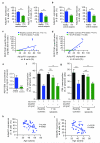
Figure 5. The PEPITEM pathway is compromised in T1D and rheumatoid arthritis
(a, b) The expression of (left) adiponectin receptor-1 or (right) adiponectin receptor-2 on B cells in cohorts of healthy controls (a: n=19; b: n=10) and in (a) individuals with T1D (n=29) (b) or rheumatoid arthritis (RA) (n=11–12). (c, d) Correlation between expression of (c) AdipoR1 and (d) AdipoR2 on B cells and the quantity of PEPITEM released by B cells following adiponectin stimulation measured by mass spectrometry in healthy controls and individuals with T1D, n=10 in each group. (e) Concentrations of PEPITEM in serum from healthy controls and individuals with T1D, n=7. (f, g) T cell transmigration in individuals (f) with T1D (n=9–22) or (g) rheumatoid arthritis (RA) (n=7–8) after treatment with adiponectin (15 μg/ml) or PEPITEM (10 ng/ml) or scrambled peptide (10 ng/ml). (h, i) Correlation between expression of (h) AdipoR1 and (i) AdipoR2 on B cells and age in healthy controls, n=40–41. NS= not significant. Data are mean±s.e.m. * P≤0.05, ** P≤0.01, *** P≤0.001 compared to (a) healthy control by Mann-Whitney test or (b, e) unpaired t-test or (f, g) paired t-test for healthy controls and (f, g) ANOVA and Dunnett post-test for individuals with T1D and rheumatoid arthritis (c, d, h, i) by linear regression.
Figure 6
Figure 6. Schematic depicting B cell-mediated regulation of T cell trafficking during inflammation
(1) Endothelial cells stimulated with pro-inflammatory cytokines such as TNF-α and IFN-γ recruit flowing T cells in the blood. (2) β1- and β2-integrins are activated by chemokine signals from CXCL9–11 presented on the endothelial surface which interact with CXCR3 on T cells leading to T cell arrest. At the same time, chemokine stimulation induces S1PR1/4 surface expression on T cells (3). T cell spreading and migration is supported by PGD2, generated through the metabolism of arachidonic acid (AA) by cyclooxygenases (COX), which operates through the PGD2 receptor, DP-2 (4) T cells spread and migrate across and through vascular endothelial cells. (5) Simultaneously, adherent B cells are simulated by circulating adiponectin through the AdipoR1/2 receptors, (6) resulting in the release of PEPITEM from B cells. (7) PEPITEM binds to Cadherin-15 (CDH15) on endothelial cells, stimulating the release of S1P from endothelial cells. (8) S1P binds to S1P receptors (S1P1/4) on recruited T cells, (9), which regulates the activation of the β2-integrin LFA-1 and inhibits trans-endothelial T cell migration.

Comment in

References

    1. Cyster JG. Lymphoid organ development and cell migration. Immunol Rev. 2003;195:5–14. - PubMed
    1. Koulmanda M, et al. Modification of adverse inflammation is required to cure new-onset type 1 diabetic hosts. Proc Natl Acad Sci U S A. 2007;104:13074–13079. - PMC - PubMed
    1. Pober JS, Cotran RS. The role of endothelial cells in inflammation. Transplantation. 1990;50:537–544. - PubMed
    1. Kadowaki T, et al. Adiponectin and adiponectin receptors in insulin resistance, diabetes, and the metabolic syndrome. J Clin Invest. 2006;116:1784–1792. - PMC - PubMed
    1. Ouedraogo R, et al. Adiponectin deficiency increases leukocyte-endothelium interactions via upregulation of endothelial cell adhesion molecules in vivo. J Clin Invest. 2007;117:1718–1726. - PMC - PubMed

METHODS-ONLY REFERENCES

    1. Rainger GE, Stone P, Morland CM, Nash GB. A novel system for investigating the ability of smooth muscle cells and fibroblasts to regulate adhesion of flowing leukocytes to endothelial cells. J Immunol Methods. 2001;255:73–82. - PubMed
    1. Cooke BM, Usami S, Perry I, Nash GB. A simplified method for culture of endothelial cells and analysis of adhesion of blood cells under conditions of flow. Microvasc Res. 1993;45:33–45. - PubMed
    1. Butler LM, Rainger GE, Rahman M, Nash GB. Prolonged culture of endothelial cells and deposition of basement membrane modify the recruitment of neutrophils. Exp Cell Res. 2005;310:22–32. - PubMed
    1. Eidhammer I, et al. Computational and Statistical Methods for Protein Quantification by Mass Spectrometry. John Wiley & Sons; New Jersey: 2012.
    1. Rajakariar R, et al. Novel biphasic role for lymphocytes revealed during resolving inflammation. Blood. 2008;111:4184–4192. - PMC - PubMed

Publication types

MeSH terms