. 2015 Jun:135:192-205.
doi: 10.1016/j.exer.2015.04.009. Epub 2015 Apr 18.

The BALB/c mouse: Effect of standard vivarium lighting on retinal pathology during aging

Affiliations

The BALB/c mouse: Effect of standard vivarium lighting on retinal pathology during aging

Brent A Bell et al. Exp Eye Res. 2015 Jun.

Abstract

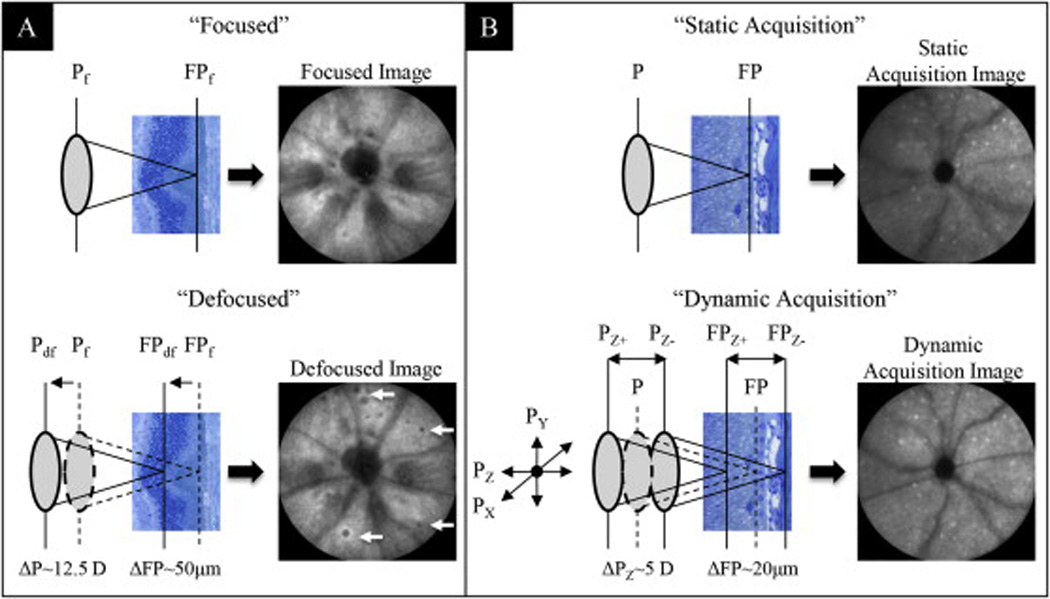
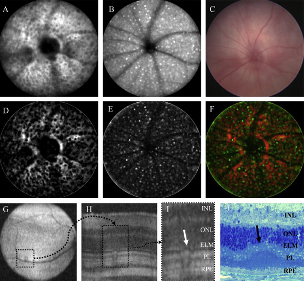
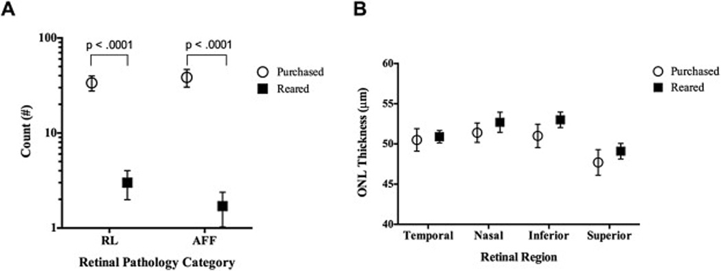
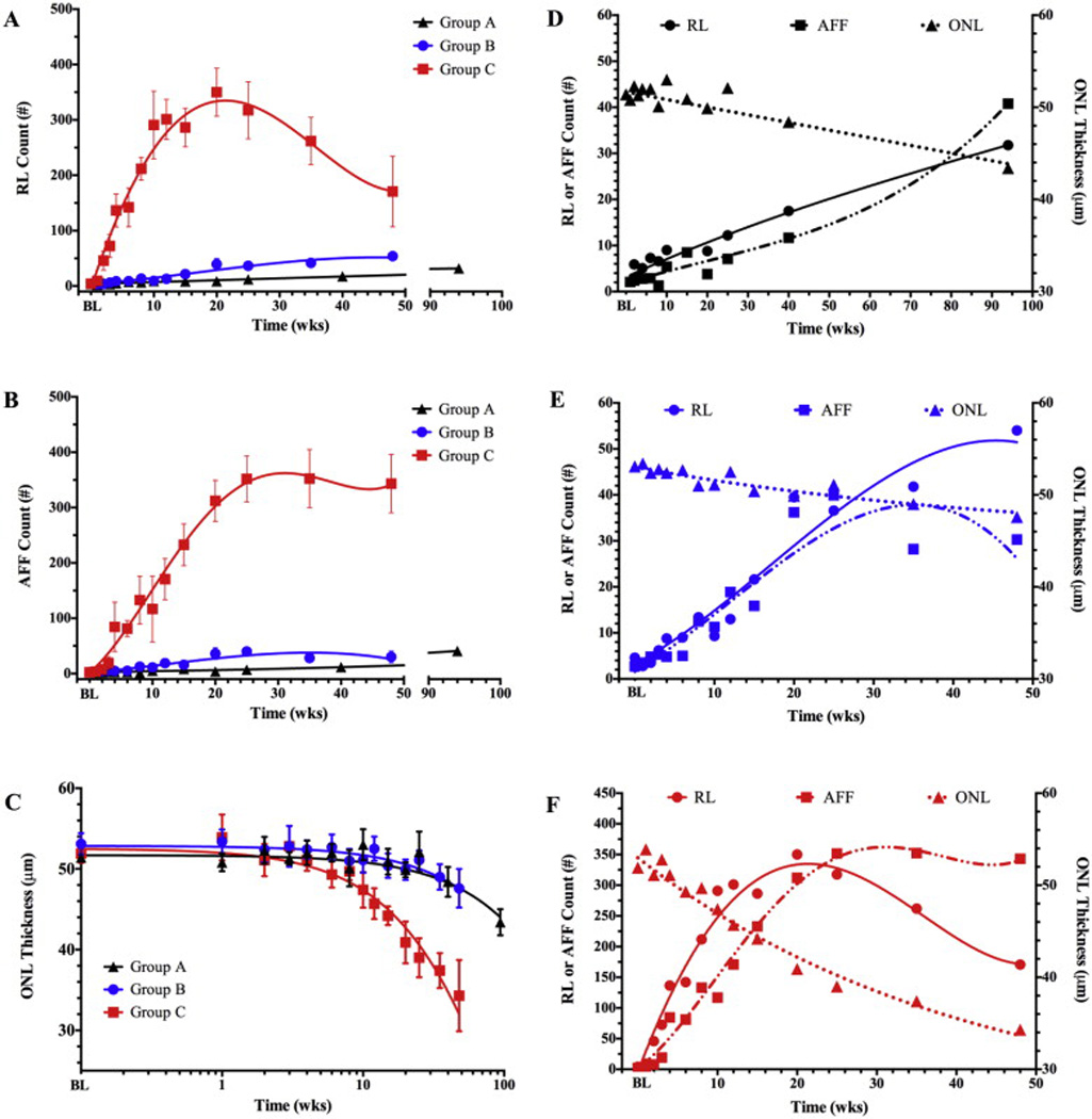
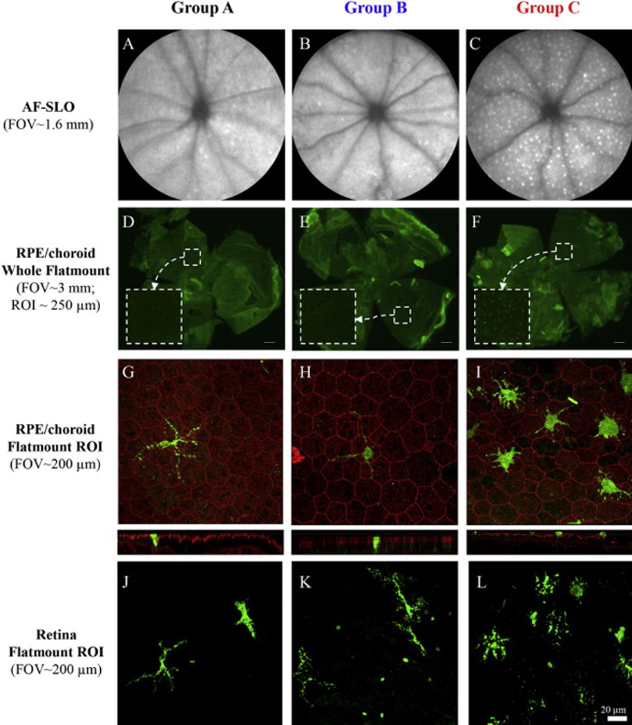
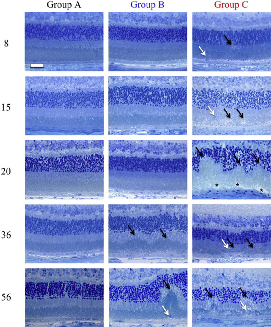
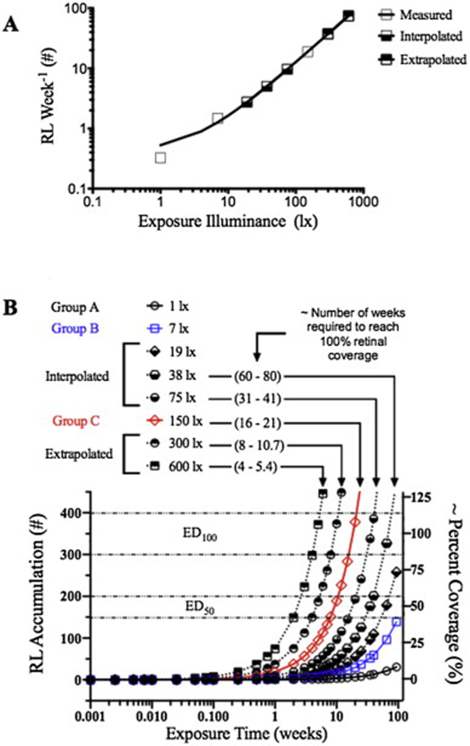
BALB/cJ mice housed under normal vivarium lighting conditions can exhibit profound retinal abnormalities, including retinal infoldings, autofluorescent inflammatory cells, and photoreceptor degeneration. To explore the sensitivity of the outer retina to cyclic lighting during aging, a cohort of BALB/cJ mice was evaluated with Scanning Laser Ophthalmoscopy (SLO), Spectral-Domain Optical Coherence Tomography (OCT) and conventional histopathology. Mice were bred and reared in a low-illuminance (extracage/intracage: 13 lx/1 lx) vivarium under cyclic light (14 h light: 10 h dark). Retinal imaging (around postnatal day 70) was performed to screen for any pre-existing abnormalities and to establish a baseline. Mice with normal retinas were separated into groups (A, B, C) and placed on bottom (Groups A & B) or top (Group C) of the cage racks where cage illumination was <10 & 150 lx respectively. Experimental groups B & C were imaged multiple times over a 17 month period. Mice from group A (controls) were imaged only once post-baseline at various times for comparison to groups B & C. Mice were assessed by histology at 8, 15, 20, 36, and 56 weeks and immunohistochemistry at 15 weeks post-baseline. SLO and OCT retinal images were measured and the resulting trends displayed as a function of age and light exposure. Retinal lesions (RL) and autofluorescent foci (AFF) were identified with histology as photoreceptor layer infoldings (IF) and localized microglia/macrophages (MM), respectively. Few RL and AFF were evident at baseline. Retinal infoldings were the earliest changes followed by subjacent punctate autofluorescent MM. The colocalization of IF and MM suggests a causal relationship. The incidence of these pathological features increased in all groups relative to baseline. OCT imaging revealed thinning of the outer nuclear layer (ONL) in all groups at 1 year relative to baseline. ONL thinning followed an exponential rate of change but the decay constant varied depending on intensity of illumination of the groups. Advanced age and top row illuminance conditions resulted in significant photoreceptor cell loss as judged by decreased thickness of the ONL. Photoreceptor loss was preceded by both retinal infoldings and the presence of autofluorescent inflammatory cells in the outer retina, suggesting that these changes are early indicators of light toxicity in the BALB/cJ mouse.

Keywords: BALB/c; Degeneration; Imaging; Inflammation; Infolding; Mice; Phototoxicity; Retina.


Figures

Fig. 1
Fig. 1
Image enhancement techniques for optimal visualization of RL (A) and AFF (B) in BALB/cJ mice. Two manual adjustment methods applied to the SLO Z-axis focus knob were used for optimization of image acquisition. Both methods enhanced the ability to detect abnormalities in the outer retina. In IR mode, focal setting adjustments from “Focused” to “Defocused” repositioned the confocal image plane from the RPE/Choroid complex to a more proximal location in the photoreceptor layer. This adjustment enhanced the ability to visualize RL (arrows). In AF mode, axial focus manipulation involved manually rocking the Z-axis (Pz) focus knob back and forth while acquiring the image with the automated realtime tracking and averaging (25 frames) function activated. This method enhanced the ability to capture an image of uniform intensity as well as improve the ability to visualize AFF (white punctate foci) in the outer retina.
Fig. 2
Fig. 2
Multimodal imaging and processing to identify retinal pathology in the BALB/cJ mouse. Fig. 2A–C is a comparison between IR-SLO (A) and AF-SLO (B) and visible light fundus (C) images after 20 weeks of exposure to 150 lx cyclic-light conditions. SLO is highly sensitive at detecting widespread changes that occur in the outer retina as a result of light stress induced by ambient vivarium lighting conditions. Visible light fundus techniques are incapable of detecting these changes due a lack of contrast between the respective abnormalities and background caused by the lack of melanopigment in the RPE-choroid complex. Fig. 2C–F demonstrates coregistration of RL and AFF abnormalities, respectively, to show that they are interrelated (F). For this comparison Fig. 2A and B were processed in ImageJ to accentuate RL (D; dark regions on a red background) and AFF (E; bright pseudocolored green foci), respectively. An image merge (F) shows the amount of overlap observed between these two abnormalities detected by different SLO imaging modes. Fig. 2G–J demonstrates detection and confirmation of RL abnormalities by non-invasive OCT imaging and histomorphology techniques. Under optimal conditions, OCT imaging is also capable of visualizing SLO identified RL lesions (G; dotted square in OCT en face image) which show altered architecture (H; dotted rectangle in OCT B-scan) of the photoreceptor layer (I; ROI). Histomicrophotograph (J) from a different mouse shows a single photoreceptor infolding (arrow). As a result of these observations from multiple assessment techniques we are confident that RL observed using SLO are retinal photoreceptor infoldings. Infoldings appear to interfere with SLO image acquisition by disrupting the light signal collection efficiency that is returned from outer retina lamina, RPE and choroidal structures. (Fig. 2I, J abbrev: INL-inner nuclear layer, ONL-outer nuclear layer, ELM-external limiting membrane, PL-photoreceptor layer, RPE-retinal pigmented epithelium).
Fig. 3
Fig. 3
SLO and OCT results from BALB/cJ mice (Age: 9 ± 1 week) obtained from a reputable vendor compared to those reared in-house. Fig. 3A shows that mice from the vendor, which also went through an unregulated (i.e. light level >> 30 lx) quarantine period upon arrival, are compromised relative to animals that are born and reared in-house under low-light level (<30 lx) conditions. Purchased mice had significantly elevated levels of both RL and AFF relative to animals bred/reared in-house. OCT results (B) showed no significant differences in ONL thickness between the purchased vs. in-house reared mice.
Fig. 4
Fig. 4
Time-dependent changes (top) in retinal pathology extracted from SLO (A, B) and OCT (C) images of BALB/cJ mice from treatment Grps A, B, & C. No significant differences in RL, AFF count or ONL thickness were observed at baseline (BL). Grp C has significant increases in RL (A) at 3 (p = .0003), 4, 6, 8, 10, 15, 20, & 25 weeks (4–25 wks; p < .0001), AFF number (B) at 4, 6, 8, 10, 15, 20, & 25 weeks (4–25 wks; p < .0001), and ONL degeneration (C) at 2 (p = .0044), 10, 15, 20, & 25 weeks (10–25 wks; p < .0001). Dynamic comparisons between RL, AFF, and ONL degeneration are individually shown for Grps A (D), B (E), & C (F). Fig. 4F shows the relationship between these three retinal observations in Grp C mice and reveals that RL count precedes AFF count and ONL degeneration by 1 and 9 weeks, respectively.
Fig. 5
Fig. 5
Representative AF-SLO images (A–C) and fluorescence microscopy of retinal flatmounts (D–F) from the 3 BALB/cJ mice study groups. Flatmount immunohistochemistry results using IBA1 antibody confirm observations made from SLO imaging which showed that AFF are more prevalent in Grp C than Grps A & B. IBA1+ cells (green) could be activated resident microglia or infiltrating macrophages and are shown against RPE background with Phalloidan labeling (red). After separation of retina from the RPE/choroid, laser scanning confocal microscopy of both retina (G–I) and RPE/choroid (J–K) flatmount components indicate IBA1+ cells. Z-stack images from these preparations indicated that the IBA1+ cells are located in close proximity to the apical surface of the RPE and within the photoreceptor layer outer segments. LSCM imaging of the retinal component also revealed IBA1+ cells with dendritic processes extending into the photoreceptor layer (J–K). Immunophenotypic morphology of IBA1+ cells are different in Group C relative to Groups A & B. Positive cells in Group C have larger, swollen cell bodies with shorter dendritic processes as compared to those present in Groups A & B. IBA1+ cellular density is much higher in Group C. (Scale bars: D-F = 200 µm, G-L = 20 µm). (For interpretation of the references to color in this figure legend, the reader is referred to the web version of this article.)
Fig. 6
Fig. 6
Representative photomicrographs (64x) of retinas from experimental treatment groups at time points of 8, 15, 20, 36, & 56 weeks. Grp A shows typical histomorphology for a BALB/cJ mouse that has been protected from extended light stress by being reared and housed in 1 lx conditions. Grp B demonstrates changes that occur as a result of being housed in 7 lx conditions which causes an increase in the spontaneous formation of IF and infiltration by IM. Profound changes (i.e. complete remodeling of photoreceptor layer) can be observed in the outer retina of mice that receive long-term exposure to 150 lx (Group C) compared to 7 lx (Group B) and 1 lx-controls (Group A). Between 8 and 20 weeks the IF (black arrows) grow until finally impacting the IS, OS, ELM and ONL layers in mice from treatment Grp C. Compromised IPM (black asterisk) and pyknotic RPE (below asterisk) are observed at 20 weeks. After ~20 weeks the IF begin to collapse and regress (black arrows) leaving attenuated and disorganized photoreceptor outer segments as well as engorged IM (white arrows). IM located within the photoreceptor layer and adjacent to the RPE can be observed in all groups as indicated by white arrows. Scale Bar (upper left) = 20 µm.
Fig. 7
Fig. 7
A model of RL development in the BALB/cJ mouse. Fig. 7A shows experimentally obtained (open boxes), as well as interpolated (bottom-filled boxes) and extrapolated (top-filled boxes) values, for the weekly count of RL that are anticipated to appear in an SLO image (with optic disk centrally located in the image frame) as a function of mean intracage light exposure level between the range of 1–600 lx. Fig. 7B transposes the data shown in Fig. 7A into a dosimetry diagram. The diagram reveals the relationship between RL accumulation, intracage light exposure intensity and exposure duration. This diagram anticipates when BALB/c mice will reach the point where the retina will have 50% (ED50) and 100% (ED100) coverage of retinal infoldings. Both of these model diagrams assume that RL growth proceeds at a linear rate between 1 and 600 lx until 100% coverage (i.e. maximum) is reached. Beyond maximum coverage the rate will plateau and/or decrease as shown in Fig. 4A-Group C. The range for ED100 was determined to be ~300–400 which is where Grp C mice in Fig. 4A reached a maximum RL count. ED50 was estimated to be 1/2 of ED100.

References

    1. Akhmedov NB, Piriev NI, Chang B, Rapoport AL, Hawes NL, Nishina PM, Nusinowitz S, Heckenlively JR, Roderick TH, Kozak CA, Danciger M, Davisson MT, Farber DB. A deletion in a photoreceptor-specific nuclear receptor mRNA causes retinal degeneration in the rd7 mouse. Proc. Natl. Acad. Sci. U. S.A. 2000;97:5551–5556. - PMC - PubMed
    1. Bell BA, Kaul C, Rayborn ME, Hollyfield JG. Baseline imaging reveals preexisting retinal abnormalities in mice. Adv. Exp. Med. Biol. 2012;723:459–469. - PubMed
    1. Bell BA, Kaul C, Hollyfield JG. A protective eye shield for prevention of media opacities during small animal ocular imaging. Exp. Eye Res. 2014;127:280–287. - PMC - PubMed
    1. Chollangi S, Wang J, Martin A, Quinn J, Ash JD. Preconditioning-induced protection from oxidative injury is mediated by leukemia inhibitory factor receptor (LIFR) and its ligands in the retina. Neurobiol. Dis. 2009;34:535–544. - PMC - PubMed
    1. Cruz-Guilloty F, Saeed AM, Echegaray JJ, Duffort S, Ballmick A, Tan Y, Betancourt M, Viteri E, Ramkhellawan GC, Ewald E, Feuer W, Huang D, Wen R, Hong L, Wang H, Laird JM, Sene A, Apte RS, Salomon RG, Hollyfield JG, Perez VL. Infiltration of proinflammatory m1 macrophages into the outer retina precedes damage in a mouse model of age-related macular degeneration. Int. J. Inflam. 2013;2013:503725. - PMC - PubMed

Publication types