Review
. 2015:166:51-83.
doi: 10.1007/978-3-319-16555-4_3.

Theranostic Magnetic Nanostructures (MNS) for Cancer

Affiliations
Review

Theranostic Magnetic Nanostructures (MNS) for Cancer

Vikas Nandwana et al. Cancer Treat Res. 2015.

Abstract

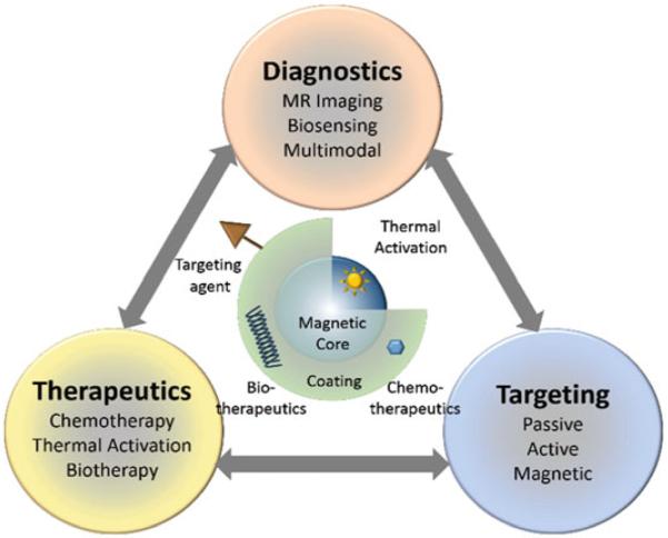
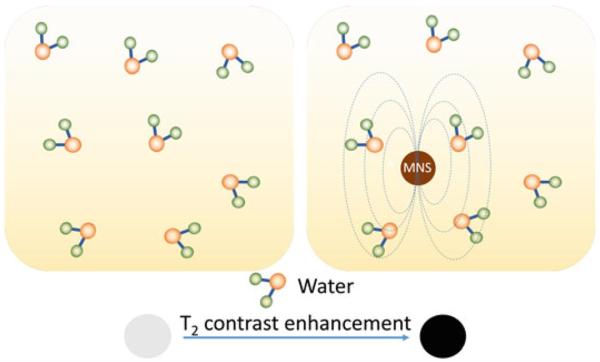
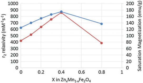
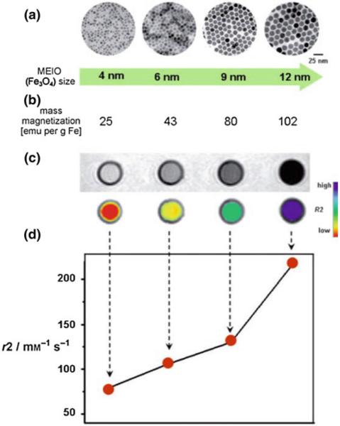
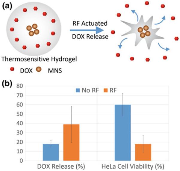
Despite the complexities of cancer, remarkable diagnostic and therapeutic advances have been made during the past decade, which include improved genetic, molecular, and nanoscale understanding of the disease. Physical science and engineering, and nanotechnology in particular, have contributed to these developments through out-of-the-box ideas and initiatives from perspectives that are far removed from classical biological and medicinal aspects of cancer. Nanostructures, in particular, are being effectively utilized in sensing/diagnostics of cancer while nanoscale carriers are able to deliver therapeutic cargo for timed and controlled release at localized tumor sites. Magnetic nanostructures (MNS) have especially attracted considerable attention of researchers to address cancer diagnostics and therapy. A significant part of the promise of MNS lies in their potential for "theranostic" applications, wherein diagnostics makes use of the enhanced localized contrast in magnetic resonance imaging (MRI) while therapy leverages the ability of MNS to heat under external radio frequency (RF) field for thermal therapy or use of thermal activation for release of therapy cargo. In this chapter, we report some of the key developments in recent years in regard to MNS as potential theranostic carriers. We describe that the r₂relaxivity of MNS can be maximized by allowing water (proton) diffusion in the vicinity of MNS by polyethylene glycol (PEG) anchoring, which also facilitates excellent fluidic stability in various media and extended in vivo circulation while maintaining high r₂values needed for T₂-weighted MRI contrast. Further, the specific absorption rate (SAR) required for thermal activation of MNS can be tailored by controlling composition and size of MNS. Together, emerging MNS show considerable promise to realize theranostic potential. We discuss that properly functionalized MNS can be designed to provide remarkable in vivo stability and accompanying pharmacokinetics exhibit organ localization that can be tailored for specific applications. In this context, even iron-based MNS show extended circulation as well as diverse organ accumulation beyond liver, which otherwise renders MNS potentially toxic to liver function. We believe that MNS, including those based on iron oxides, have entered a renaissance era where intelligent synthesis, functionalization, stabilization, and targeting provide ample evidence for applications in localized cancer theranostics.


Figures

Fig. 1
Fig. 1
Size scale of MNS as compared to biomolecules. MNS can be adapted to include biomolecules, drugs, or targeting and imaging molecules to form targeted MNS theranostic agents
Fig. 2
Fig. 2
Functional architecture of MNS and theranostic modalities. MNS are comprised of thermally active magnetic core and biocompatible coating and/or functionalization that allows integration of targeting agents and bio/chemotherapeutics
Fig. 3
Fig. 3
T2 contrast enhancement in water due to MNS. When water molecules diffuse into the periphery of the induced dipole moment by MNS, the T2 relaxation time of the water protons is shortened which enhances the negative contrast
Fig. 4
Fig. 4
MFe2O4 (where M = Mn, Fe, Co, Ni) MNS with inverse spinel structure and its magnetic spin alignments. The mass magnetization values and r2 relaxivity values of MFe2O4 MNS are proportional to the magnetic moments of the divalent ions (M2+) [38]
Fig. 5
Fig. 5
Saturation magnetization and r2 relaxivity (at 4.5 T) of (ZnxMn1–x)Fe2O4 MNS at different Zn2+ doping levels. The (ZnxMn1–x)Fe2O4 MNS showed significantly high r2 relaxivities compared to conventional iron oxide MNS [42]
Fig. 6
Fig. 6
Schematic illustration of RES clearance of MNS. MNS larger than 100 nm are absorbed by circulating opsonin proteins that are recognized by macrophages and removed from the bloodstream
Fig. 7
Fig. 7
Passive targeting of MNS via enhanced permeability and retention (EPR) effect. The compromised vasculature of a solid tumor facilitates extravasation of MNS of size less than 200 nm from the circulation into the tumor interstitium, while endothelial cells are closely packed and present a barrier for MNS penetration
Fig. 8
Fig. 8
Size effects of Fe3O4 MNS on r2 relaxivity. a TEM images, b saturation magnetization values, c T2-weighted MR images (top black and white, bottom color), and d the r2 relaxivity values of 4, 6, 9, and 12 nm sized Fe3O4 MNS. The r2 relaxivity value increased with size of Fe3O4 MNS which resulted in the T2 contrast change from light gray to black in T2 weighted MR images or from red to blue in the corresponding color-coded images. Reprinted with permission from [27]. Copyright 2005 American Chemical Society
Fig. 9
Fig. 9
In vivo MR detection of cancer in a mouse implanted with the cancer cell line NIH3T6.7 using 12 nm MnFe2O4, 12 nm Fe3O4 and dextran coated cross-linked 4 nm Fe3O4 (CLIO) MNS. T2-weighted MR images of the mouse (a) before injection, (b) after 1 h injection, and (c) after 2 h injection of MnFe2O4 in comparison to (d) after 2 h injection of CLIO. MnFe2O4-Herceptin conjugates produced higher contrast than CLIO-Herceptin conjugates at the tumor site after 2 h. e Plot of R2 change versus time. Increase in R2 up to 34 % was observed for MnFe2O4-Herceptin conjugates in comparison to 5 and 13 % for CLIO-Herceptin conjugate (dots) and 12 nm Fe3O4-Herceptin conjugates, respectively. f Change in R2 values was confirmed in the ex vivo MR images of explanted tumors (8 h). Reprinted with permission from [38]. Copyright 2007 Nature Publishing Group
Fig. 10
Fig. 10
MR signal enhancement by assembly of Fe3O4 on SiO2 nanoparticles. a Schematic illustration of the synthetic procedure for Fe3O4 decorated mesoporous silica nanoparticles. b Relaxivity values and T2 weighted MR image of Fe3O4 decorated SiO2 nanoparticles (Fe3O4-MSN) and free Fe3O4 nanoparticles. The r2 relaxivity of Fe3O4 decorated SiO2 nanoparticles was increased by 2.8 times as compared to free Fe3O4 nanoparticles, hence darker signal was observed in T2 weighted MR image at the same concentration of Fe. Reprinted with permission from Ref. [150]. Copyright 2010 American Chemical Society
Fig. 11
Fig. 11
r2 relaxivity values of 12 nm nitrodopamine-PEG functionalized Fe3O4 MNS with molecular weight of PEG 200 (EG2), 400 (EG4), 500 (EG5), and 600 Da (EG6) in comparison to Ferumoxytol and Ferumoxides (unpublished). It was found that the PEG coating not only provides stability but the thickness of PEG coating also affects the r2 relaxivity of Fe3O4 MNS. The highest r value of 396 mM−1 s−1 with PEG 600 (EG6) was almost four times that of Feridex (Ferumoxide)
Fig. 12
Fig. 12
SAR values and percentage of HeLa cells killed after treatment with (Zn0.4Mn0.6)Fe2O4 MNS or Feridex in AMF. Fluorescence microscopy images of HeLa cells treated with (Zn0.4Mn0.6) Fe2O4 nanoparticles (or Feridex) and stained with calcein show live cells as green fluorescence. (Zn0.4Mn0.6)Fe2O4 MNS have shown SAR value of 432 W/g, ~4 times higher than SAR of Feridex (115 W/g) which resulted in 84.4 % death of HeLa cancer cells in comparison to 13.5 % from Feridex. Reprinted with permission from Ref. [42] Copyright © 2009 WILEY-VCH Verlag GmbH & Co. KGaA, Weinheim
Fig. 13
Fig. 13
a Schematic illustration of drug (DOX) release from thermoresponsive hydrogel-MNS composite. b Percent DOX release and cell viability of HeLa cell lines treated with the hydrogel-MNS composite with and without external RF field [83]

References

    1. Kim J, Piao Y, Hyeon T. Multifunctional nanostructured materials for multimodal imaging, and simultaneous imaging and therapy. Chem Soc Rev. 2009;38(2):372–390. - PubMed
    1. Sun C, Lee JSH, Zhang MQ. Magnetic nanoparticles in MR imaging and drug delivery. Adv Drug Deliv Rev. 2008;60(11):1252–1265. - PMC - PubMed
    1. Ho D, Sun XL, Sun SH. Monodisperse magnetic nanoparticles for theranostic applications. Acc Chem Res. 2011;44(10):875–882. - PMC - PubMed
    1. Yoo D, et al. Theranostic magnetic nanoparticles. Acc Chem Res. 2011;44(10):863–874. - PubMed
    1. Veiseh O, Gunn JW, Zhang MQ. Design and fabrication of magnetic nanoparticles for targeted drug delivery and imaging. Adv Drug Deliv Rev. 2010;62(3):284–304. - PMC - PubMed

LinkOut - more resources