Review
. 2015 May;13(5):255-68.
doi: 10.1038/nrmicro3433.

Living in the matrix: assembly and control of Vibrio cholerae biofilms

Affiliations
Review

Living in the matrix: assembly and control of Vibrio cholerae biofilms

Jennifer K Teschler et al. Nat Rev Microbiol. 2015 May.

Abstract

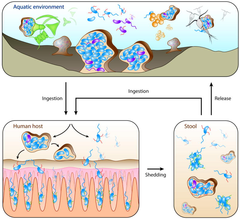
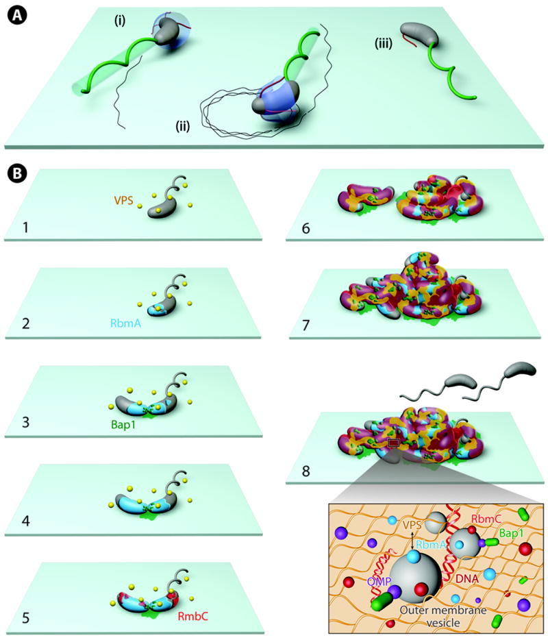
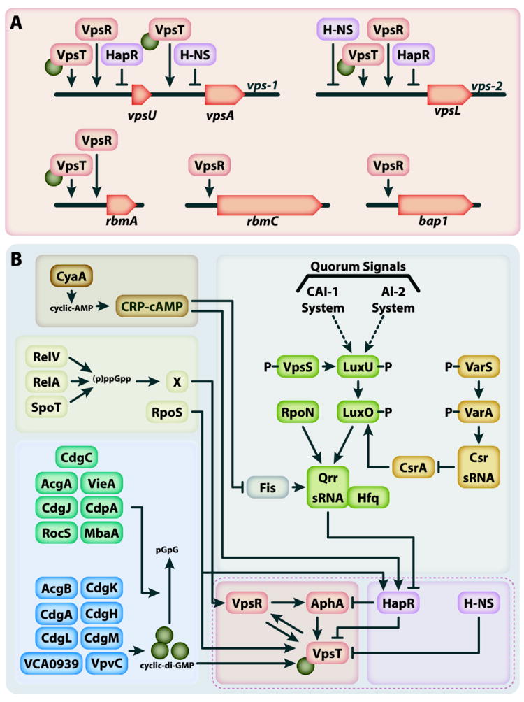
Nearly all bacteria form biofilms as a strategy for survival and persistence. Biofilms are associated with biotic and abiotic surfaces and are composed of aggregates of cells that are encased by a self-produced or acquired extracellular matrix. Vibrio cholerae has been studied as a model organism for understanding biofilm formation in environmental pathogens, as it spends much of its life cycle outside of the human host in the aquatic environment. Given the important role of biofilm formation in the V. cholerae life cycle, the molecular mechanisms underlying this process and the signals that trigger biofilm assembly or dispersal have been areas of intense investigation over the past 20 years. In this Review, we discuss V. cholerae surface attachment, various matrix components and the regulatory networks controlling biofilm formation.


Figures

Figure 1
Figure 1. Biofilms in V. cholerae life cycle
In the aquatic environment V. cholerae is found in its highly mobile planktonic form as well as in biofilms formed on zooplankton, phytoplankton, detritus, and other surfaces, such as sediments. Following the initial stages of attachment to abiotic and biotic surfaces, which involves the type IV pili mannose-sensitive haemagglutinin (MSHA) pili, cells produce the extracellular matrix, which is essential to achieve mature biofilms with a three-dimensional structure. Because it is unknown whether the flagellum is lost during biofilm formation, cells are depicted with or without the flagellum in biofilms. V. cholerae can be ingested by humans from environmental sources causing seasonal outbreaks. During intestinal colonization, V. cholerae produce toxin co-regulated pili (TCP). Both planktonic cells and biofilm aggregates are found in patient stool, and these cells can re-infect a new host or return to the aquatic environment.
Figure 2
Figure 2. Building a V. cholerae biofilm
A) Surface motility and initial attachment: Surface-skimming cells use flagella to move and mechanically ‘scan’ the surface via mannose-sensitive haemagglutinin (MSHA) pili appendages. Weak interactions between surfaces and pili lead to ‘roaming’ behavior (tight, repetitive, near-circular orbits with high curvatures), whereas strong surface-pili interactions lead to ‘orbiting’ behavior (long directional persistence and small curvatures), which allow cells to loiter over these regions and eventually attach and initiate microcolony formation. Motility trajectories are depicted by dashed lines on the surface and correspond to roaming and orbiting behavior. B) Microcolony formation and matrix production. Soon after initial attachment, Vibrio polysaccharide (VPS) is excreted from cell surfaces (B1), and VPS extrusion is observed throughout biofilm formation. Next, the biofilm matrix protein RbmA accumulates on the cell surface (B2). During cell division, the daughter cell remains attached to the founder cell (also known as parental cell), confirming the role of RbmA in cell-cell adhesion, and the biofilm matrix protein Bap1 is excreted between the two cells and on the substrate near the two cells (B3). Bap1 gradually radially accumulates on nearby surfaces, although the concentration of Bap1 remains the highest near the founder cell. Subsequently, the biofilm matrix protein RbmC is excreted and found on discrete sites on the cell surface (B4). As biofilms develop, VPS, RbmC and Bap1 form envelopes that can grow as cells divide (B5). The mature biofilm is a composite of organized clusters composed of cells, VPS, RbmA, Bap1 and RbmC, in addition to other matrix components, such as outer membrane vesicles (OMVs) and extracellular DNA (eDNA) (inset). Outer membrane proteins (OMPs) associate with Bap1 in OMVs and bind to antimicrobial peptides thereby increasing V. cholerae resistance. The last stages in biofilm development are dispersal, whereby exiting V. cholerae cells seek out and colonize new resources (B6); however, the underlying mechanism remains to be determined.
Figure 3
Figure 3. V. cholerae biofilm regulatory network
A) The transcriptional activators VpsR, VpsT and transcriptional repressors HapR and H-NS directly and indirectly regulate several genes that have key roles in biofilm formation. Positive regulators of biofilm are shown in orange, while negative regulators are shown in purple. These include the vps (vibrio polysaccharide) cluster and the rbm (rugosity and biofilm structure modulator) cluster, which contain genes that encode proteins involved in VPS production and matrix proteins. The vps and rbm clusters comprise a functional genetic module, the V. cholerae biofilm-matrix cluster (VcBMC). In addition, the bap1 (biofilm-associated extracellular matrix protein) gene has also been shown to be regulated by these core regulators. The recognition sequences for VpsR, VpsT, HapR and H-NS have been identified in the regulatory region of vps-1 and vps-2 clusters and the genes encoding the extracellular matrix proteins RbmA and RbmC. Binding of VpsT to promoter regions requires its interaction with c-di-GMP. As shown, the VpsR and VpsT targets extensively overlap, though some biofilm genes appear to be only directly regulated by one. Additionally, the negative regulators directly downregulate many of the genes encoding proteins involved in VPS production and matrix proteins, as well as the genes that encode the positive transcriptional regulators of those genes (shown in part b). B) An extensive regulatory network governs V. cholerae biofilm formation. VpsR, VpsT and AphA are the main activators of biofilm formation, and HapR and H-NS are the main repressors (shown in the core the dashed box). VpsR, VpsT, HapR and H-NS directly regulate genes involved in biofilm formation (see part a). These core regulators directly and indirectly regulate each other and are modulated by a complex regulatory network in response to a number of environmental and host signals. The quorum sensing (QS) pathway, which responds to cell density via bacterial signaling, has a key role in the regulation of HapR and, thus, the other major biofilm regulators. The signaling molecules autoinducer 2 (AI-2) and cholerae autoinducer 1 (CAI-1) regulate a phosphorelay event that culminates at the histidine phosphotransfer protein, LuxU, and the response regulator, LuxO. Together with the alternative sigma factor RpoN, LuxO activates transcription of the quorum-regulated small RNAs (sRNAs), Qrr1–4, which work in conjunction with the sRNA chaperone Hfq to prevent the translation of hapR. HapR production is repressed at low cell density, when CAI-1 and AI-2 production is not high, shown by dashed arrows, leading to LuxO phosphorylation by the QS signal transduction pathway. The VarS-VarA system responds to an unknown environmental cue and represses biofilm production by post-transcriptionally upregulating HapR. This process involves the regulatory sRNAs CsrB, CsrC, and CsrD, which bind to and titrate the RNA-binding protein CsrA, thereby interfering with LuxO-mediated activation of Qrr1-4. This leads to decreased levels of Qrr1-4 and enhanced HapR production. By contrast, the small protein Fis is a direct positive regulator of the QS-responsive sRNAs, Qrr1-4 thereby promoting HapR repression. The histidine kinase VpsS, donates phosphate groups to LuxU, thus promoting HapR repression. The integration of many regulatory pathways enables the induction or repression of V. cholerae biofilm formation in response to a number of extracellular and intracellular signals. Small-nucleotide molecules, including cyclic-AMP (cAMP), (p)ppGpp, and cyclic-di-GMP regulate the induction and repression of major regulators, including HapR, VpsT and VpsR. The sigma factor RpoS promotes expression of hapR. Of note, RpoS is depicted with (p)ppGpp because the stringent response regulation of vpsT and vpsR has been shown to partially occur through RpoS. A key signaling molecule controlling V. cholerae motility and biofilm matrix production is the second messenger c-di-GMP. High cellular levels of c-di-GMP promote enhanced transcription of genes involved in biofilm formation, possibly by promoting VpsT-mediated transcriptional expression of vps genes. Several diguanylate cyclases (DGCs), which cumulatively contribute to c-di-GMP levels, and phosphodiesterases (PDEs), known to degrade cellular c-di-GMP to pGpG or GMP, are shown. The second messenger cyclic adenosine-monophosphate (cAMP) is involved various cellular responses and acts as a repressor of V. cholerae biofilm formation. cAMP in complex with cAMP receptor protein (CRP), has been shown to upregulate HapR production through its positive regulation of the CAI-I autoinducer synthase and its negative regulation of Fis. Finally, the sigma factor RpoS promotes expression of hapR.
Figure
Figure
Figure
Figure

Similar articles

Cited by

References

    1. Charles RC, Ryan ET. Cholera in the 21st century. Curr Opin Infect Dis. 2011;24:472–7. - PubMed
    1. Islam MS, et al. Biofilm acts as a microenvironment for plankton-associated Vibrio cholerae in the aquatic environment of Bangladesh. Microbiol Immunol. 2007;51:369–79. - PubMed
    1. Faruque SM, et al. Transmissibility of cholera: in vivo-formed biofilms and their relationship to infectivity and persistence in the environment. Proc Natl Acad Sci U S A. 2006;103:6350–5. This study demonstrates that the ingestion of metabolically quiescent V. cholerae cells can lead to the formation of in vivo biofilms that are hyperinfective when excreted in patient stool. - PMC - PubMed
    1. Alam M, et al. Viable but nonculturable Vibrio cholerae O1 in biofilms in the aquatic environment and their role in cholera transmission. Proc Natl Acad Sci U S A. 2007;104:17801–6. This study reveals the crucial role of biofilms in maintaining environmental reservoirs between epidemics, as well as the role of host passage in activation of metabolically quiescent cells. - PMC - PubMed
    1. Matz C, et al. Biofilm formation and phenotypic variation enhance predation-driven persistence of Vibrio cholerae. Proc Natl Acad Sci U S A. 2005;102:16819–24. - PMC - PubMed

Publication types

MeSH terms