. 2015 Jul;83(7):2636-50.
doi: 10.1128/IAI.00025-15.

The Serine Protease Autotransporter Pic Modulates Citrobacter rodentium Pathogenesis and Its Innate Recognition by the Host

The Serine Protease Autotransporter Pic Modulates Citrobacter rodentium Pathogenesis and Its Innate Recognition by the Host

Kirandeep Bhullar et al. Infect Immun. 2015 Jul.

Abstract

Bacterial pathogens produce a number of autotransporters that possess diverse functions. These include the family of serine protease autotransporters of Enterobacteriaceae (SPATEs) produced by enteric pathogens such as Shigella flexneri and enteroaggregative Escherichia coli. Of these SPATEs, one termed "protein involved in colonization," or Pic, has been shown to possess mucinase activity in vitro, but to date, its role in in vivo enteric pathogenesis is unknown. Testing a pic null (ΔpicC) mutant in Citrobacter rodentium, a natural mouse pathogen, found that the C. rodentium ΔpicC strain was impaired in its ability to degrade mucin in vitro compared to the wild type. Upon infection of mice, the ΔpicC mutant exhibited a hypervirulent phenotype with dramatically heavier pathogen burdens found in intestinal crypts. ΔpicC mutant-infected mice suffered greater barrier disruption and more severe colitis and weight loss, necessitating their euthanization between 10 and 14 days postinfection. Notably, the virulence of the ΔpicC mutant was normalized when the picC gene was restored; however, a PicC point mutant causing loss of mucinase activity did not replicate the ΔpicC phenotype. Exploring other aspects of PicC function, the ΔpicC mutant was found to aggregate to higher levels in vivo than wild-type C. rodentium. Moreover, unlike the wild type, the C. rodentium ΔpicC mutant had a red, dry, and rough (RDAR) morphology in vitro and showed increased activation of the innate receptor Toll-like receptor 2 (TLR2). Interestingly, the C. rodentium ΔpicC mutant caused a degree of pathology similar to that of wild-type C. rodentium when infecting TLR2-deficient mice, showing that despite its mucinase activity, PicC's major role in vivo may be to limit C. rodentium's stimulation of the host's innate immune system.


Figures

FIG 1
FIG 1
(A) Characterization of mucinase activity of picC in C. rodentium. Zones of mucin (bovine submaxillary mucin [BSM]) clearance are visible in the stacking region of the SDS-PAGE gel (boxed area), indicative of mucinase activity. Deletion of picC (the C. rodentium ΔpicC mutant) results in loss of mucinase activity, which is restored after complementation of picC (C. rodentium ΔpicC + pPicC). Incubation of samples with PMSF significantly reduced mucinase activity, with the exception of the C. rodentium ΔpicC mutant (no mucinase activity). (B) Reduced mucinase activity also was seen with a C. rodentium strain expressing a PicC protein containing a mutation in the serine-protease active site, S258I. Lanes labeled BSM represent untreated mucin. The gels are stained with PAS.
FIG 2
FIG 2
C57BL/6 mice exhibit dramatic susceptibility to the C. rodentium ΔpicC mutant. (A) Weight loss of WT- and ΔpicC mutant-infected mice, plotted as a percentage of initial body weight and normalized to day 0 body weight. Error bars represent SEM, and asterisks indicate significant differences (***, P < 0.0005) by the Mann-Whitney test. (B) Survival curve of C57BL/6 mice following WT and ΔpicC mutant infection. P values (0.0253) are from the log-rank test and indicate a statistically significant difference between the survival curves. Results are representative of 3 independent experiments (12 mice per group).
FIG 3
FIG 3
C. rodentium ΔpicC mutant-infected mice carry heavier pathogen burdens. (A) Adherent (distal and cecal tissues) and nonadherent luminal C. rodentium burdens at 8 dpi. (B) Representative images from distal colon showing C. rodentium localization as seen via Tir (C. rodentium-specific effector; red) and DAPI (counterstain; blue) staining. Original magnification, ×200. WT C. rodentium is seen mostly on the epithelial surface (inset, top row, ×630), whereas the C. rodentium ΔpicC mutant penetrates deeper into the crypts (inset, bottom row, ×630). (C) Quantitative analysis looking at Tir-positive crypts/100 crypts. Analysis was done at the original magnification, ×200, and represents averages for distal colons from at least 9 different mice/group. Error bars represent SEM, and asterisks indicate significant difference (*, P < 0.05; **, P < 0.01; ***, P < 0.0005) by the Mann-Whitney test.
FIG 4
FIG 4
Heightened histopathological damage and increased proinflammatory cytokines in C. rodentium ΔpicC mutant-infected mice. (A) Representative H&E-stained distal colon (original magnification, ×100) at 8 dpi for WT- and ΔpicC mutant-infected mice. Asterisks represent damage to the intestinal epithelial surface, as seen by ruffling and loss of crypt structure and/or epithelial integrity. Arrows point to increased edema and infiltration of immune/inflammatory cells. (B) Cumulative tissue pathology damage scores from distal colon of WT- and ΔpicC mutant-infected mice. Scoring was done by blinded observers and represents averages from 9 mice/group. (C) Quantitative PCR of proinflammatory cytokine and chemokine genes in the distal colon of the WT- and ΔpicC mutant-infected mice at 8 dpi. Note that WT C. rodentium infection resulted in increased gene transcript levels of tested chemokines and cytokines; however, there was a greater induction seen with C. rodentium ΔpicC mutant infection, indicative of hyperinflammatory responses. Errors bars represent SEM from three independent experiments and at least 9 mice/group. P < 0.01 (**) and P < 0.05 (*) by the Mann-Whitey test.
FIG 5
FIG 5
C. rodentium ΔpicC mutant-infected mice have impaired epithelial barrier integrity and increased translocation of pathogenic and commensal bacteria. (A) C. rodentium ΔpicC mutant-infected mice display greater FITC-dextran flux across their intestinal barrier on 6 dpi than WT C. rodentium-infected mice. The bar graph shows the quantity of FD4 in serum and represents an average of 9 mice/group. UN, uninfected C57BL/6 mice. (B) Quantification of C. rodentium burdens recovered from systemic sites (liver, spleen, and MLN) on 8 dpi. (C) Recovery of viable commensal bacteria from MLN under controlled anaerobic conditions and commensal-specific media. Errors bars represent SEM from 3 independent experiments. P < 0.0005 (***), P < 0.01 (**), and P < 0.05 (*) by the Mann-Whitney test.
FIG 6
FIG 6
Mucinase activity of PicC is not essential for intestinal colonization. (A) C. rodentium burdens enumerated at 8 dpi from systemic sites, i.e., liver, spleen, and MLN, for WT C. rodentium- and S258I C. rodentium (catalytic mutant with no mucinase activity)-infected mice. (B) WT and S258I mutant burdens recovered from distal colon, cecum, and lumen at 8 dpi. (C) Representative H&E-stained distal colon (original magnification, ×100) at 8 dpi. (D) Histologic damage scores from distal colon of WT- and S258I mutant-infected mice. Pathology scoring included submucosal edema, hyperplasia, goblet cell depletion, damage to epithelial integrity, and PMN infiltration. Scores were determined by 2 independent observers under blinded conditions. (E) FITC-dextran intestinal permeability assay for WT- and S258I mutant-infected C57BL/6 mice. The baseline FITC-dextran flux for uninfected C57BL/6 mice also is shown. Uninfected and infected mice were gavaged with FITC-dextran, and serum was collected using cardiac puncture. Serum FD4 levels were measured. Asterisks represent statistically significant differences (*, P < 0.05 by the Mann-Whitney test); ns, not significant. Errors bars show SEM from 3 independent infections and 9 mice/group.
FIG 7
FIG 7
C. rodentium PicC is not a potent mucus secretagogue in vivo. (A) Representative dual immunofluorescence staining in Carnoy's fixed distal colon at 8 dpi. Mucin Muc2 (green), Tir (red; shows the association of C. rodentium with Muc2-positive crypts), and DAPI (nuclei; counterstain) are shown. The white arrowhead indicates secreted/luminal mucus, and the yellow arrowhead shows representative Muc2-filled goblet cells. (B) Representative PAS/AB staining of Carnoy's fixed distal colon collected from WT- and ΔpicC mutant-infected mice. Arrows point to the secreted mucus. (C) Quantification of total mucus secretion in WT- and ΔpicC mutant-infected mice at 6 dpi. Plot shows the counts per minute (cpm) in individual column fractions containing radioactive 3H activity after total secretions were subjected to gel filtration chromatography (Sepharose 4B column). This plot represents averages from 6 mice/group and 2 independent infections. The inset shows a bar graph representation of total counts per minute in void (Vo) volume fractions (points 10 to 20, large, mucin glycoproteins) plotted as a percentage of uninfected samples. Vt represents fractions eluting nonmucin glycoproteins.
FIG 8
FIG 8
C. rodentium ΔpicC mutant forms microcolony-like structures in vivo. (A) Representative Muc2/Tir dual staining profile in distal colon. White arrowheads show localization of WT C. rodentium mostly on the epithelial surface in a single layer. Yellow arrowheads point to clustering of the C. rodentium ΔpicC mutant close to epithelial surfaces and within the colonic crypts. Original magnification, ×630. Blue, DAPI counterstain. (B) Dual FISH staining in distal colon using EUB338 DNA probe (stains all bacteria red) and GAM42 DNA probe (stains Gammaproteobacteria green; C. rodentium belongs to this family). Yellow, C. rodentium; red, commensals; L, lumen. White arrows point to microcolony-like structures on the mucosal surface in C. rodentium ΔpicC mutant sections not seen in WT C. rodentium sections. Original magnification, ×200. The last panel is a magnified image (×630) of the white boxed area showing a microcolony intermixed with commensals and the C. rodentium ΔpicC mutant. (C) RDAR colony morphology of WT C. rodentium (top) and the C. rodentium ΔpicC mutant (bottom) on Congo red plates incubated at 25°C for 5 days. Note the differences in colony morphology between WT C. rodentium and the C. rodentium ΔpicC mutant and the dry, rough phenotype localized in the central region of the C. rodentium ΔpicC mutant colonies.
FIG 9
FIG 9
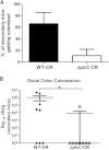
C. rodentium ΔpicC mutant shows reduced transmission to new hosts. (A) Bar graph showing percent transmission (i.e., percentage of naive secondary mice getting colonized from exposure to index mice infected with the WT or C. rodentium ΔpicC mutant). The C. rodentium ΔpicC mutant displayed reduced transmission to new hosts compared to WT C. rodentium. (B) Viable C. rodentium (CFU) recovered from distal colon of secondary mice. Note that index mice were mixed with naive (secondary) mice on 6 dpi and cohoused for 48 h, after which all mice were euthanized. Results shown are representative of 2 independent infections. The asterisk indicates statistical significance (*, P < 0.05 by the Mann-Whitney test).
FIG 10
FIG 10
PicC plays a role in innate immune recognition through TLR2. (A and B) TLR reporter assay. HEK-Blue hTLR4 (A) and HEK-Blue hTLR2 (B) were exposed to supernatants from either live or heat-killed (HK) C. rodentium WT and ΔpicC mutant for 4 h. The C. rodentium ΔpicC mutant significantly increased the stimulation of TLR2 compared to WT C. rodentium. HK bacteria maxed out the TLR4 and TLR2 stimulation under tested conditions. Error bars indicate SEM. P < 0.0005 (***) and P < 0.01 (**) by Mann-Whitney test; ns, not significant. Values represent the means from three independent experiments. (C) Body weights of Tlr2−/− mice infected with WT C. rodentium and the C. rodentium ΔpicC mutant, plotted as a percentage of starting body weight. (D) H&E-stained representative images showing histological damage to distal colon of WT- and ΔpicC mutant-infected Tlr2−/− mice at 8 dpi (original magnification, ×200). (E) Pathology scores comparing damage to distal colon in Tlr2−/− mice infected with WT C. rodentium and the C. rodentium ΔpicC mutant. Each bar represents an average score from 6 tissues, scored under blinded conditions. Error bars represent SEM, and results from 3 independent experiments are pooled.

References

    1. Wales AD, Woodward MJ, Pearson GR. 2005. Attaching-effacing bacteria in animals. J Comp Pathol 132:1–26. doi:10.1016/j.jcpa.2004.09.005. - DOI - PMC - PubMed
    1. Luperchio SA, Schauer DB. 2001. Molecular pathogenesis of Citrobacter rodentium and transmissible murine colonic hyperplasia. Microbes Infect 3:333–340. doi:10.1016/S1286-4579(01)01387-9. - DOI - PubMed
    1. Mundy R, MacDonald TT, Dougan G, Frankel G, Wiles S. 2005. Citrobacter rodentium of mice and man. Cell Microbiol 7:1697–1706. doi:10.1111/j.1462-5822.2005.00625.x. - DOI - PubMed
    1. Bry L, Brenner MB. 2004. Critical role of T cell-dependent serum antibody, but not the gut-associated lymphoid tissue, for surviving acute mucosal infection with Citrobacter rodentium, an attaching and effacing pathogen. J Immunol 172:433–441. doi:10.4049/jimmunol.172.1.433. - DOI - PubMed
    1. Gibson DL, Ma C, Rosenberger CM, Bergstrom KS, Valdez Y, Huang JT, Khan MA, Vallance BA. 2008. Toll-like receptor 2 plays a critical role in maintaining mucosal integrity during Citrobacter rodentium-induced colitis. Cell Microbiol 10:388–403. doi:10.1111/j.1462-5822.2007.01052.x. - DOI - PubMed

Publication types

MeSH terms