. 2015 Jul;83(7):2651-60.
doi: 10.1128/IAI.03083-14. Epub 2015 Apr 20.

AP-1 Transcription Factor Serves as a Molecular Switch between Chlamydia pneumoniae Replication and Persistence

Affiliations

AP-1 Transcription Factor Serves as a Molecular Switch between Chlamydia pneumoniae Replication and Persistence

S Krämer et al. Infect Immun. 2015 Jul.

Abstract

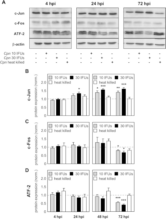
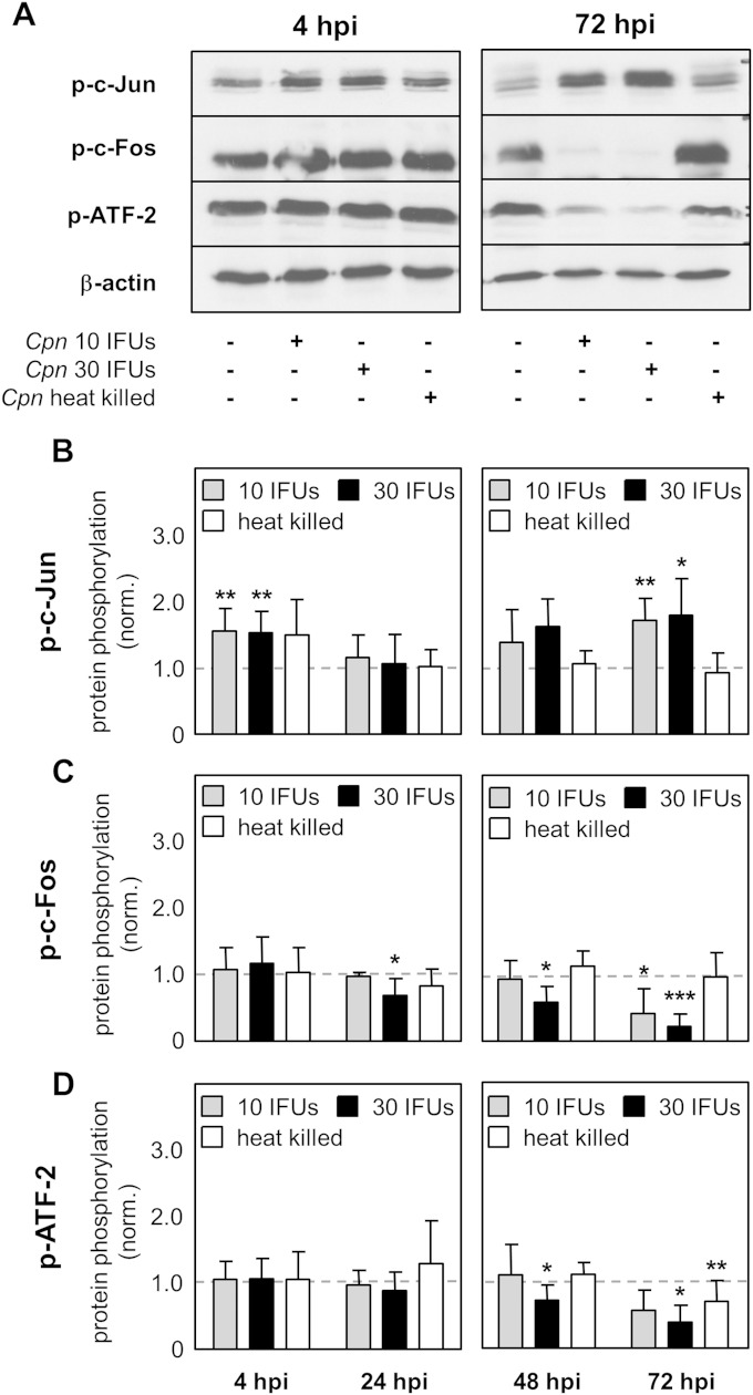
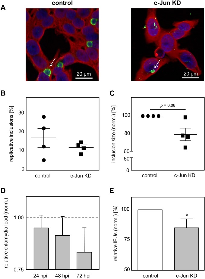
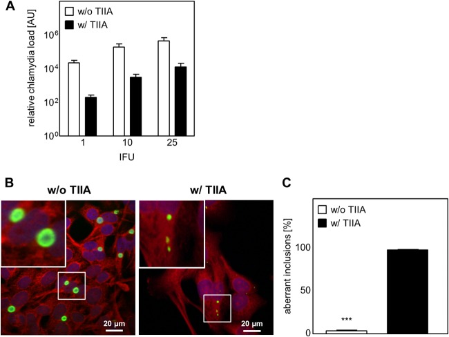
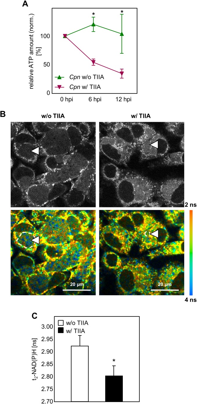
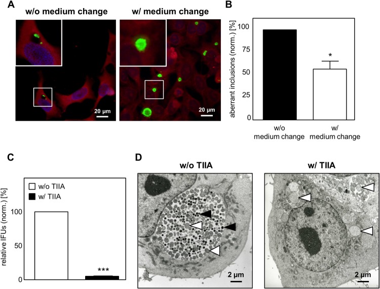
Chlamydia pneumoniae is a Gram-negative bacterium that causes acute or chronic respiratory infections. As obligate intracellular pathogens, chlamydiae efficiently manipulate host cell processes to ensure their intracellular development. Here we focused on the interaction of chlamydiae with the host cell transcription factor activator protein 1 (AP-1) and its consequence on chlamydial development. During Chlamydia pneumoniae infection, the expression and activity of AP-1 family proteins c-Jun, c-Fos, and ATF-2 were regulated in a time- and dose-dependent manner. We observed that the c-Jun protein and its phosphorylation level significantly increased during C. pneumoniae development. Small interfering RNA knockdown of the c-Jun protein in HEp-2 cells reduced the chlamydial load, resulting in smaller inclusions and significantly lower chlamydial recovery. Furthermore, inhibition of the c-Jun-containing AP-1 complexes using tanshinone IIA changed the replicative infection phenotype into a persistent one. Tanshinone IIA-dependent persistence was characterized by smaller, aberrant inclusions, a strong decrease in the chlamydial load, and significantly reduced chlamydial recovery, as well as by the reversibility of the reduced recovery after the removal of tanshinone IIA. Interestingly, not only was tanshinone IIA treatment accompanied by a significant decrease of ATP levels, but fluorescence live cell imaging analysis by two-photon microscopy revealed that tanshinone IIA treatment also resulted in a decreased fluorescence lifetime of protein-bound NAD(P)H inside the chlamydial inclusion, indicating that chlamydial reticulate bodies have decreased metabolic activity. In all, these data demonstrate that the AP-1 transcription factor is involved in C. pneumoniae development, with tanshinone IIA treatment resulting in persistence.


Figures

FIG 1
FIG 1
Differential regulation of AP-1 proteins after C. pneumoniae infection. Lysates from uninfected HEp-2 cells (control), HEp-2 cells infected with 10 IFUs (low dose) or 30 IFUs (high dose) of C. pneumoniae (Cpn), or HEp-2 cells infected with heat-killed C. pneumoniae were prepared at various time points after infection (4 hpi, 24 hpi, 48 hpi, and 72 hpi). (A) Western blot of the indicated proteins at 4 hpi, 24 hpi, and 72 hpi. (B to D) Using densitometry, expression of the c-Jun (B), c-Fos (C), and ATF-2 (D) proteins was quantified during C. pneumoniae infection of HEp-2 cells. Data were normalized (norm.) against those for the uninfected control (dashed gray lines). Data, presented as the mean ± SD, and immunoblots are representative of those from at least four independent experiments (n = 4 to 6). *, P < 0.05; **, P < 0.01; ***, P < 0.001.
FIG 2
FIG 2
Phosphorylation of AP-1 proteins after C. pneumoniae infection. Lysates from uninfected HEp-2 cells (control), HEp-2 cells infected with 10 IFUs (low dose) or 30 IFUs (high dose) of C. pneumoniae (Cpn), or HEp-2 cells infected with heat-killed C. pneumoniae were prepared at various time points after infection (4 hpi, 24 hpi, 48 hpi, and 72 hpi). (A) The phosphorylation of the c-Jun, c-Fos, and ATF-2 proteins and the expression of β-actin were analyzed by Western blot analysis at the indicated time points (4 hpi and 72 hpi). (B to D) Using densitometry, phosphorylation of the c-Jun (B), c-Fos (C), and ATF-2 (D) proteins was quantified during C. pneumoniae infection of HEp-2 cells. Data were normalized against those for the uninfected control (dashed gray lines). Data are presented as the mean ± SD. Data and immunoblots are representative of those from at least four independent experiments (n = 4 to 6). p-c-Jun, p-c-Fos, and p-ATF-2, phosphorylated c-Jun, c-Fos, and ATF-2, respectively. *, P < 0.05; **, P < 0.01; ***, P < 0.001.
FIG 3
FIG 3
Knockdown of the c-Jun protein impairs C. pneumoniae infection. (A) Infected cells with and without c-Jun knockdown (c-Jun KD) were methanol fixed at 48 hpi, immunostained by use of an Imagen chlamydia kit (green, chlamydial LPS; red, cytoplasm), and counterstained with DAPI (blue). White arrows, C. pneumoniae inclusions. (B and C) From the immunofluorescence images, the amount of pathological inclusions (B) and the size (C) of control and c-Jun-KD cells were quantified during infection. (D) Quantification of the relative C. pneumoniae load in c-Jun KD cells using qRT-PCR. (E) Chlamydial recovery after infection with C. pneumoniae cells obtained from infected control or c-Jun-specific siRNA-treated cells. The data in panels C to E were normalized against those for the control (dashed gray line in panel D). Data represent the mean ± SD. Data, immunoblots, and immunofluorescence images are representative of those from at least three independent experiments (n = 3 to 7). *, P < 0.05.
FIG 4
FIG 4
Tanshinone IIA treatment decreases the chlamydial load and limits inclusion development. HEp-2 cells were infected with C. pneumoniae (1, 10, or 25 IFUs) and in parallel treated with (w/) or without (w/o) tanshinone IIA (TIIA; 25 μM). After 48 h the cells were harvested, followed by mRNA isolation. (A) Quantification of relative chlamydial load using qRT-PCR. AU, arbitrary units. (B) Infected cells (10 IFUs) with and without tanshinone IIA treatment were fixed with methanol after 48 h, immunostained by use of an Imagen chlamydia kit (green, chlamydial LPS; red, cytoplasm), and counterstained with DAPI (blue). (Insets) Magnified images of the boxed areas. (C) Aberrant inclusions were quantified in C. pneumoniae-infected cells with and without tanshinone IIA treatment. Data, presented as the mean ± SD, and immunofluorescence images are representative of those from at least three independent experiments (n = 3 to 4). ***, P < 0.001.
FIG 5
FIG 5
The ATP level and protein-bound NAD(P)H fluorescence lifetime decreased after tanshinone IIA treatment. (A) Quantification of the relative amount of ATP. HEp-2 cells were infected with 10 IFUs of C. pneumoniae (Cpn) and treated with or without tanshinone IIA (TIIA; 25 μM). Relative ATP levels were measured directly (0 hpi) and at 6 hpi and 12 hpi. (B and C) HEp-2 cells were infected with C. pneumoniae, and tanshinone IIA was applied in parallel, followed by incubation for 48 h. (B) Gray-scale images of the NAD(P)H fluorescence intensity (top) and color-coded images of the protein-bound NAD(P)H fluorescence lifetime (τ2-NAD(P)H) (bottom) in C. pneumoniae-infected cells not treated (left) and treated (right) with tanshinone IIA. Dashed lines and arrowheads indicate representative chlamydial inclusions which were selected for quantitative analysis. ns, nanoseconds. (C) Quantitative analysis of τ2-NAD(P)H inside the chlamydial inclusion. Chlamydial inclusions (n = 30) from three different samples were analyzed on two independent experimental days. Data, presented as the mean ± SEM, are representative of those from three independent experiments (n = 3). *, P < 0.05.
FIG 6
FIG 6
Inhibition of AP-1 (c-Jun/c-Fos) transcription activity results in persistent C. pneumoniae infection. HEp-2 cells were infected with C. pneumoniae (10 IFUs), and tanshinone IIA (TIIA; 25 μM) was applied in parallel, followed by incubation for 48 h. Cells were cultured with or without tanshinone IIA for an additional 48 h. (A) Cells were fixed with methanol, immunostained by use of an Imagen chlamydia kit (green, Chlamydia LPS; red, cytoplasm), and counterstained with DAPI (blue). (Insets) Magnified images of the boxed areas. (B) Quantification of aberrant inclusions during infection without and with a medium change. (C) Chlamydial recovery 48 h after infection with and without tanshinone IIA treatment. (D) Electron microscope micrographs of infected HEp-2 cells without and with tanshinone IIA treatment. Black arrowheads, elementary bodies; white arrowheads, reticular bodies. Data represent the mean ± SD from at least three independent experiments. Immunofluorescence and electron microscope images are representative of those from three independent experiments (n = 3). *, P < 0.05; ***, P < 0.001.

Similar articles

Cited by

References

    1. Koh WP, Taylor MB, Hughes K, Chew SK, Fong CW, Phoon MC. 2002. Seroprevalence of IgG antibodies against Chlamydia pneumoniae in Chinese, Malays and Asian Indians in Singapore. Int J Epidemiol 31:1001–1007. doi:10.1093/ije/31.5.1001. - DOI - PubMed
    1. Scidmore-Carlson MA, Shaw EI, Dooley CA, Fischer ER, Hackstadt T. 1999. Identification and characterization of a Chlamydia trachomatis early operon encoding four novel inclusion membrane proteins. Mol Microbiol 33:753–765. doi:10.1046/j.1365-2958.1999.01523.x. - DOI - PubMed
    1. Harper A, Pogson CI, Jones ML, Pearce JH. 2000. Chlamydial development is adversely affected by minor changes in amino acid supply, blood plasma amino acid levels, and glucose deprivation. Infect Immun 68:1457–1464. doi:10.1128/IAI.68.3.1457-1464.2000. - DOI - PMC - PubMed
    1. Nelson DE, Virok DP, Wood H, Roshick C, Johnson RM, Whitmire WM, Crane DD, Steele-Mortimer O, Kari L, McClarty G, Caldwell HD. 2005. Chlamydial IFN-gamma immune evasion is linked to host infection tropism. Proc Natl Acad Sci U S A 102:10658–10663. doi:10.1073/pnas.0504198102. - DOI - PMC - PubMed
    1. Hogan RJ, Mathews SA, Mukhopadhyay S, Summersgill JT, Timms P. 2004. Chlamydial persistence: beyond the biphasic paradigm. Infect Immun 72:1843–1855. doi:10.1128/IAI.72.4.1843-1855.2004. - DOI - PMC - PubMed

Publication types

MeSH terms

Substances