. 2015 Jul;83(7):2725-37.
doi: 10.1128/IAI.00376-15. Epub 2015 Apr 20.

An Invertron-Like Linear Plasmid Mediates Intracellular Survival and Virulence in Bovine Isolates of Rhodococcus equi

Affiliations

An Invertron-Like Linear Plasmid Mediates Intracellular Survival and Virulence in Bovine Isolates of Rhodococcus equi

Ana Valero-Rello et al. Infect Immun. 2015 Jul.

Abstract

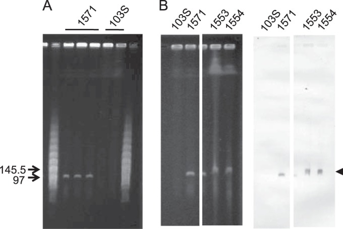
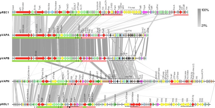
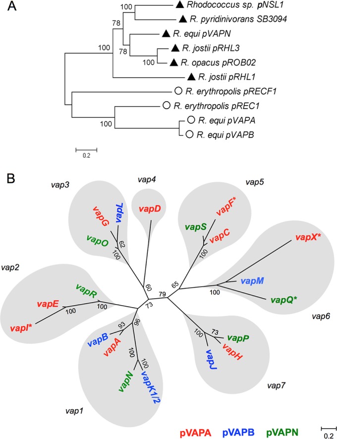
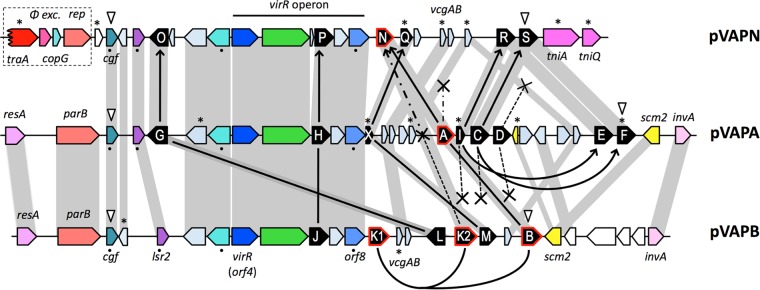
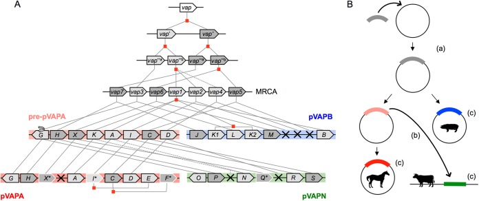
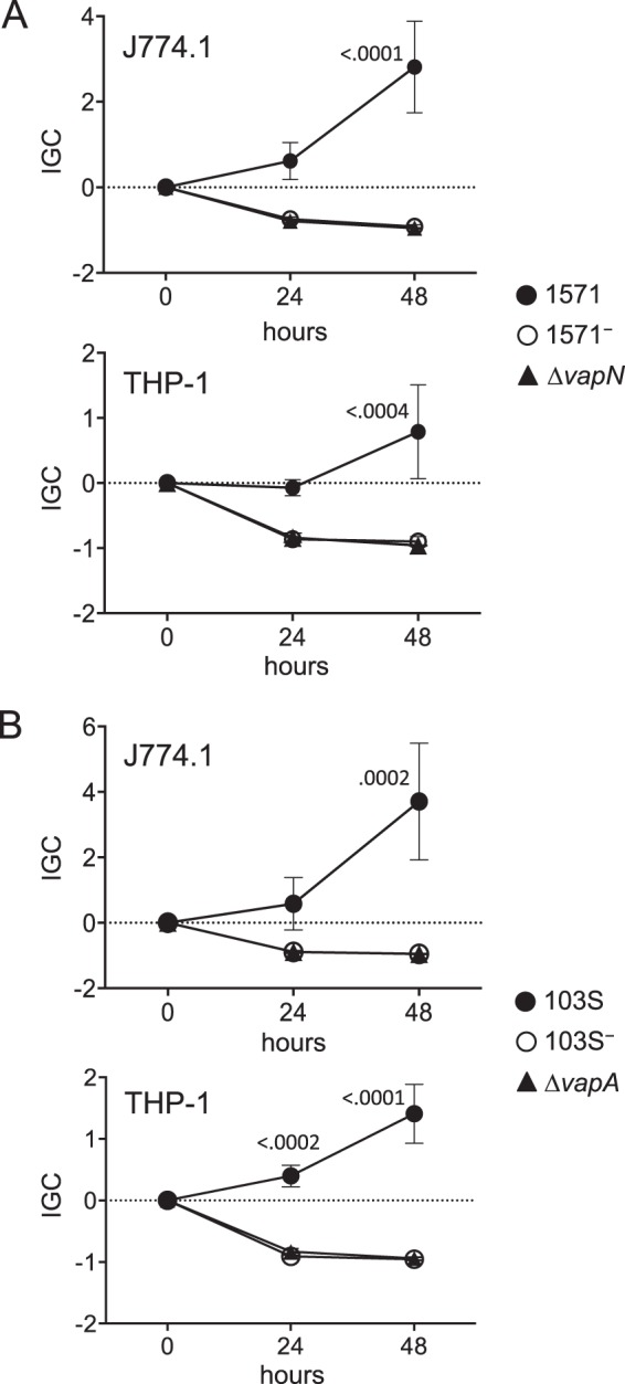
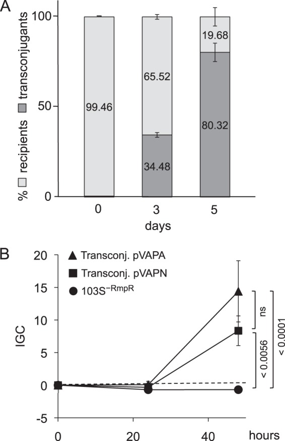
We report a novel host-associated virulence plasmid in Rhodococcus equi, pVAPN, carried by bovine isolates of this facultative intracellular pathogenic actinomycete. Surprisingly, pVAPN is a 120-kb invertron-like linear replicon unrelated to the circular virulence plasmids associated with equine (pVAPA) and porcine (pVAPB variant) R. equi isolates. pVAPN is similar to the linear plasmid pNSL1 from Rhodococcus sp. NS1 and harbors six new vap multigene family members (vapN to vapS) in a vap pathogenicity locus presumably acquired via en bloc mobilization from a direct predecessor of equine pVAPA. Loss of pVAPN rendered R. equi avirulent in macrophages and mice. Mating experiments using an in vivo transconjugant selection strategy demonstrated that pVAPN transfer is sufficient to confer virulence to a plasmid-cured R. equi recipient. Phylogenetic analyses assigned the vap multigene family complement from pVAPN, pVAPA, and pVAPB to seven monophyletic clades, each containing plasmid type-specific allelic variants of a precursor vap gene carried by the nearest vap island ancestor. Deletion of vapN, the predicted "bovine-type" allelic counterpart of vapA, essential for virulence in pVAPA, abrogated pVAPN-mediated intramacrophage proliferation and virulence in mice. Our findings support a model in which R. equi virulence is conferred by host-adapted plasmids. Their central role is mediating intracellular proliferation in macrophages, promoted by a key vap determinant present in the common ancestor of the plasmid-specific vap islands, with host tropism as a secondary trait selected during coevolution with specific animal species.


Figures

FIG 1
FIG 1
Detection of pVAPN by PFGE. (A) Genomic DNA of bovine isolate 1571 and equine isolate 103S; three and two independent lysates per strain are shown. Relevant positions of the lambda PFGE marker (New England BioLabs) are indicated. pVAPN is observable as a distinct band of ≈100 kb in the bovine isolate. (B) Southern blot analysis of bovine isolates PAM1571, PAM1533, and PAM1554 (strain 103S was used as a negative control). (Left) Relevant sections of the PFGE gel; (right) membrane hybridized with a pVAPN-specific DNA probe (600-bp fragment encompassing the 3′ region of vapN and the 5′ region of vapQ). The arrow indicates the pVAPN band.
FIG 2
FIG 2
Genome alignments of the linear virulence plasmid pVAPN, circular virulence plasmids pVAPA and pVAPB, and the respective closest homologs from nonpathogenic rhodococcal species (pNSL1 from Rhodococcus sp. NS1 [48] and pREC1 from R. erythropolis [44]). The alignments were built with EasyFig (http://easyfig.sourceforge.net/). The circular plasmids (pVAPA, pVAPB, and pREC1) were linearized starting from the first conserved gene of the housekeeping backbone. Regions with significant similarity between plasmids are connected by gray stripes (tblastx E value threshold of 0.1); grayscale indicates percent similarity. ORFs are color coded as follows: hypothetical proteins (gray), conjugation or DNA replication/recombination/metabolism (red), DNA mobility genes (magenta), transcriptional regulators (blue), secreted proteins (dark green), membrane proteins (pale green), metabolic functions (yellow), vap family genes (black), and pseudogenes (brown). Green and pale red bars below the genes indicate conjugation and replication/partitioning functional modules, respectively; dashed underline indicates HGT regions identified by Alien_hunter (52); and the triangle indicates a putative origin of replication. Abbreviations: 3oxoACPr, 3-oxoacyl-ACP reductase; 3oxoACPs, 3-oxoacyl-ACP synthase; Endo, endonuclease; Exo, exonuclease; Lig, ligase; LT, lytic transglycosylase; MT, methyltransferase; Mtase, methylase; PK, protein kinase; PR, pentapeptide repeat protein; ssRec, site-specific recombinase; TPR, TPR repeat protein; FA met, fatty acid metabolism.
FIG 3
FIG 3
ML trees of Rhodococcus plasmid backbones (A) and the R. equi vap multigene family (B). The Hasegawa-Kishino-Yano with gamma distribution (HKY+G) evolutionary model was used. (A) ML tree based on a concatenated alignment of orthologs from a selection of rhodococcal extrachromosomal replicons (total of 7,802 nucleotides). The genes used are indicated by dots in Fig. 2. Values >50 for 100 bootstrap replicates are indicated. Symbols: triangles, linear plasmids; circles, circular plasmids. (B) vap family members derived from each of the predicted seven precursor vap genes in the MRCA of the extant pVAPA, pVAPB, and pVAPN PAIs are in gray balloons.
FIG 4
FIG 4
pVAPN telomeric sequences. (A) ClustalΩ alignment of the left- and right-end 200 terminal nucleotides. Identical nucleotides are shaded (dark and light blue, purines and pyrimidines, respectively). Inverted repeats are indicated above the sequence. In red are four conserved palindromic sequences with the central motif GCTNCGC identified in the binding site of telomere-associated proteins involved in Streptomyces linear plasmid replication (72). Several “GCTNCGC” palindromic sequences are normally present in the telomeres of rhodococcal linear plasmids (43–45) (see Fig. S2 in the supplemental material). (B) Secondary structures potentially formed by the palindromic sequences in pVAPN telomeres, as numbered in panel A. Structures were determined with mFold. Free energy (ΔG) values: −33.84 kcal/mol (left), −37.95 kcal/mol (right).
FIG 5
FIG 5
Genetic structure of the vap PAIs from pVAPN (15.1 kb), pVAPA (21.5 kb), and pVAPB (15.9 kb). Color codes of genes: vap family (black), DNA conjugation/partitioning (red), DNA mobility/recombination (magenta), transcriptional regulators (blue), other regulators (cyan), membrane proteins (green), metabolic reactions (yellow). Orthologs are in the same color shade and linked by gray bands. ORFs encoding hypothetical proteins are represented in light blue-gray and in white if they are outside the PAI. White arrowheads point to the first and last genes of the consensus PAI. The traA pseudogene/phage excisionase-rep-copG HGT cluster presumably acquired by pVAPN from the pVAPA backbone is boxed. The figure also schematizes the probable evolutionary relationships of the vap multigene family as inferred from phylogenetic analyses (Fig. 3B; see also Fig. S4 and S5 in the supplemental material) and PAI genetic structure; the model minimizes the number of vap gene loss events. Solid lines/arrows connect vap genes belonging to the same monophyletic group (thus likely representing allelic variants of a common vap gene ancestor). Curved lines/arrows indicate vap gene duplications within a PAI. Crosses denote vap genes that were lost, and asterisks indicate pseudogenes. Two alternative evolutionary paths are shown for vapA-vapB-vapK1-vapK2-vapN (see the legend of Fig. S5 in the supplemental material for additional details). The black dots indicate the non-vap genes used for the MLSA shown in Fig. S4B in the supplemental material.
FIG 6
FIG 6
Hypothetical reconstruction of vap PAI evolution. (A) Model of vap multigene family evolution. Lines indicate the evolutionary path of the vap genes between ancestral PAIs, the most recent common ancestor (MRCA), and extant PAIs. Pre-pVAPA designates the hypothetical direct precursor of the current pVAPA PAI. Gene duplication events are indicated by red squares, gene loss events by crosses, and pseudogenes by asterisks and white borders. (B) Fate of the vap PAI and R. equi virulence plasmid evolution. (a) Acquisition by rhodococcal circular replicon of vap PAI ancestor conferring the ability to colonize macrophages; (b) mobilization of vap PAI from pre-pVAPA plasmid to rhodococcal linear replicon; (c) evolution of species specificity.
FIG 7
FIG 7
Intracellular proliferation in murine (J774A.1) and human (THP-1) macrophages. Data are expressed as normalized IGC values (see Materials and Methods). Means of data from three duplicate experiments ± standard errors are shown. Statistical significance was analyzed by 2-way ANOVA; P values determined by Šidák post hoc multiple-comparison tests at each time point are shown if ≤0.05. (A) Plasmidless derivative and in-frame ΔvapN mutant of bovine isolate 1571. Two-way ANOVA P values = 0.0007 for J774A.1 and 0.0160 for THP-1. (B) Plasmidless derivative and in-frame ΔvapA mutant of equine isolate 103S. Two-way ANOVA P values = 0.0112 for J774A.1 cells and <0.0001 for THP-1 cells.
FIG 8
FIG 8
Competitive virulence assay in mouse lung. BALB/c mice (n = 4 per time point) were infected intranasally with a ≈1:1 mixture of the test bacteria, and the competing populations were monitored 60 min after infection (t = 0) and then on four consecutive days. Bar height denotes total lung CFU, and the light and dark gray areas within bars indicate the proportions of the competing bacteria. Corresponding CI values are shown in Table 1. (A) Competition between wild-type bovine isolate 1571 and the isogenic plasmidless derivative 1571 at an infection dose of 3.7 × 10⁷ CFU/mouse (2.3 × 10⁷ and 1.4 × 10⁷ CFU, respectively). (B) Competition between the avirulent 1571 strain and an in-frame 1571ΔvapN deletion mutant at an infection dose of 7.8 × 10⁷ CFU/mouse (3.2 × 10⁷ and 4.6 × 10⁷ CFU, respectively).
FIG 9
FIG 9
Transfer of pVAPN by mating confers virulence to a plasmid-negative R. equi recipient strain. (A) In vivo selection of pVAPN transconjugants in mice. Note the progressive enrichment of the recipient 103S−RmpR strain upon acquisition of the pVAPN plasmid. Time zero is 60 min after infection. (B) Intracellular proliferation in J774A.1 macrophages. Acquisition of pVAPN (and control pVAPA) promotes intracellular proliferation in the recipient 103S−RmpR strain. Data are expressed as normalized IGC values (see Materials and Methods). Means of data from three duplicate experiments ± standard errors; P values (determined by 2-way ANOVA and Šidák post hoc multiple-comparison tests) are indicated. ns, not significant.

References

    1. Prescott JF. 1991. Rhodococcus equi: an animal and human pathogen. Clin Microbiol Rev 4:20–34. - PMC - PubMed
    1. Vazquez-Boland JA, Giguère S, Hapeshi A, MacArthur I, Anastasi E, Valero-Rello A. 2013. Rhodococcus equi: the many facets of a pathogenic actinomycete. Vet Microbiol 167:9–33. doi: 10.1016/j.vetmic.2013.06.016. - DOI - PubMed
    1. Muscatello G, Leadon DP, Klayt M, Ocampo-Sosa A, Lewis DA, Fogarty U, Buckley T, Gilkerson JR, Meijer WG, Vazquez-Boland JA. 2007. Rhodococcus equi infection in foals: the science of ‘rattles’. Equine Vet J 39:470–478. doi: 10.2746/042516407X209217. - DOI - PubMed
    1. Cohen ND, Chaffin MK, Kuskie KR, Syndergaard MK, Blodgett GP, Takai S. 2013. Association of perinatal exposure to airborne Rhodococcus equi with risk of pneumonia caused by R. equi in foals. Am J Vet Res 74:102–109. doi: 10.2460/ajvr.74.1.102. - DOI - PubMed
    1. Giguère S, Cohen ND, Chaffin MK, Hines SA, Hondalus MK, Prescott JF, Slovis NM. 2011. Rhodococcus equi: clinical manifestations, virulence, and immunity. J Vet Intern Med 25:1221–1230. doi: 10.1111/j.1939-1676.2011.00804.x. - DOI - PubMed

Publication types

MeSH terms

Associated data